. 2023 Jun 3;45(6):4875-4890.
doi: 10.3390/cimb45060310.

Magnolia kobus Extract Suppresses Porphyromonas gingivalis LPS-Induced Proinflammatory Cytokine and MMP Expression in HGF-1 Cells and Regulates Osteoclastogenesis in RANKL-Stimulated RAW264.7 Cells

Affiliations

Magnolia kobus Extract Suppresses Porphyromonas gingivalis LPS-Induced Proinflammatory Cytokine and MMP Expression in HGF-1 Cells and Regulates Osteoclastogenesis in RANKL-Stimulated RAW264.7 Cells

Hae Jin Lee et al. Curr Issues Mol Biol. .

Abstract

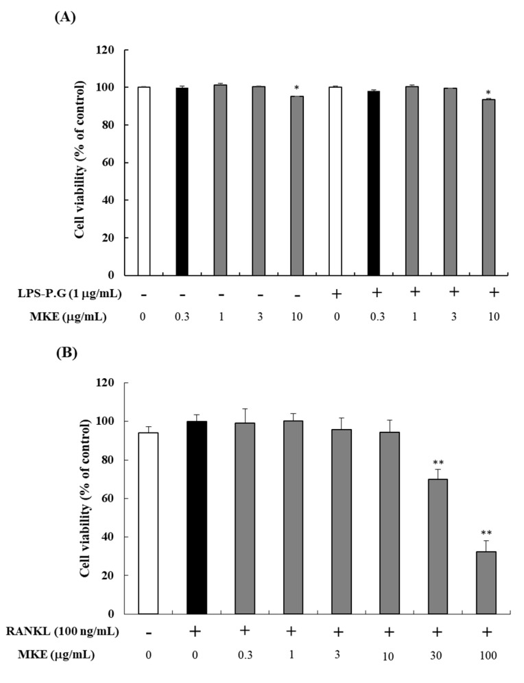
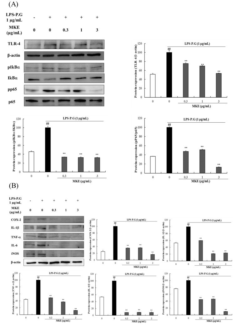
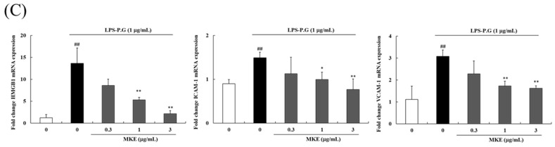
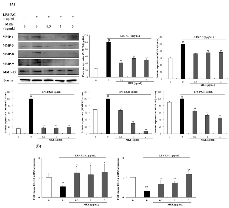
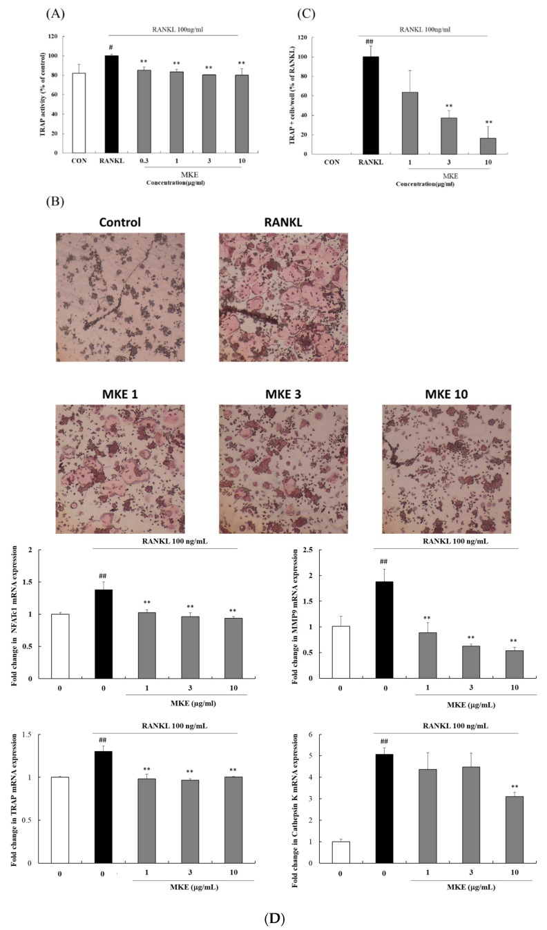
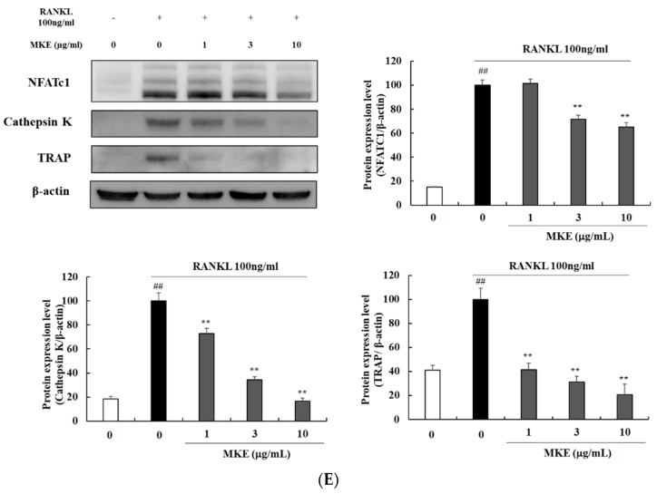
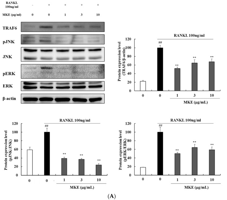
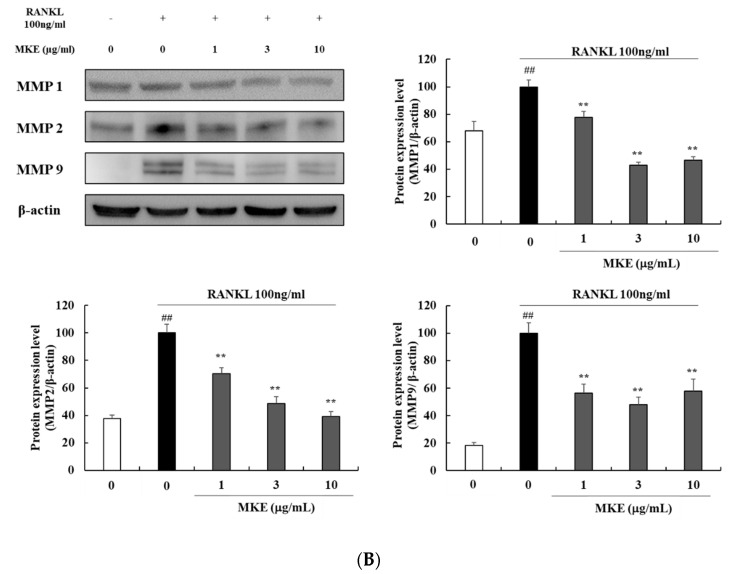
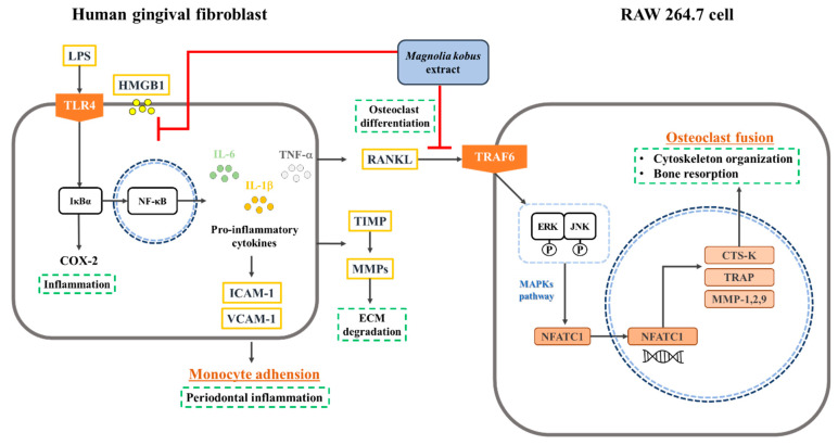
Clinical prevention is of utmost importance for the management of periodontal diseases. Periodontal disease starts with an inflammatory response in the gingival tissue, and results in alveolar bone destruction and subsequent tooth loss. This study aimed to confirm the anti-periodontitis effects of MKE. To confirm this, we studied its mechanism of action using qPCR and WB in LPS-treated HGF-1 cells and RANKL-induced osteoclasts. We found that MKE suppressed proinflammatory cytokine protein expression by inhibiting the TLR4/NF-κB pathway in LPS-PG-induced HGF-1 cells and blocking ECM degradation by regulating the expression of TIMPs and MMPs. We also confirmed that TRAP activity and multinucleated cell formation were reduced in RANKL-stimulated osteoclasts after exposure to MKE. These results were confirmed by inhibiting TRAF6/MAPK expression, which led to the suppression of NFATc1, CTSK, TRAP, and MMP expression at the gene and protein levels. Our results confirmed that MKE is a promising candidate for the management of periodontal disease based on its anti-inflammatory effects and inhibition of ECM degradation and osteoclastogenesis.

Keywords: ECM degradation; Magnolia kobus; bone resorption; gingival tissue destruction; human gingival fibroblast; inflammation; osteoclast; periodontitis.


Conflict of interest statement

Author H.J.L., S.J.L., S.K.L., B.K.C., D.R.L. was employed by the company NUON Co. The remaining authors declare that the research was conducted in the absence of any commercial or financial relationships that could be construed as a potential conflict of interest.

Figures

Figure 1
Figure 1
(A) Effects of Magnolia kobus extract (MKE) treatment (0.3, 1, 3, or 10 μg/mL) on HGF-1 cell viability. (B) Effects of MKE treatment (0, 0.3, 1, 3, 10, 30, or 100 μg/mL) on RAW264.7 cell viability. HGF-1 cells were incubated with MKE for 24 h with or without LPS-PG (1 μg/mL), while RAW264.7 cells were maintained with RANKL (100 ng/mL) for 5 days. After incubation, the cell viability was assessed using an MTT assay. The experiments (n = 3) were conducted three times independently and the values are expressed as mean ± standard deviation. * p < 0.05 and ** p < 0.01 vs. the MKE 0 μg/mL group.
Figure 2
Figure 2
Effects of Magnolia kobus extract (MKE) treatment on the expression of the (A) TLR4/NF-κB pathways, (B) inflammatory cytokines (COX-2, IL-1β, TNF-α, IL-6, and iNOS), and (C) the mRNA levels of HMGB1, ICAM-1 and VCAM-1 in 1 μg/mL LPS-PG-stimulated HGF-1 cells. HGF-1 cells were incubated in the presence of LPS-PG (1 μg/mL) and MKE (0.3, 1, or 3 μg/mL) for 15 min or 24 h. The mRNA levels were assessed via RT-PCR and protein levels were detected using a Western blot assay. The protein band density was determined using ImageJ software. Each experiment was repeated three times (n = 3) and each value was normalized to that of β-actin and GAPDH. Data are represented as mean ± standard deviation. ## p < 0.01 vs. the vehicle control group; * p < 0.05 and ** p < 0.01 vs. the LPS-PG control group.
Figure 2
Figure 2
Effects of Magnolia kobus extract (MKE) treatment on the expression of the (A) TLR4/NF-κB pathways, (B) inflammatory cytokines (COX-2, IL-1β, TNF-α, IL-6, and iNOS), and (C) the mRNA levels of HMGB1, ICAM-1 and VCAM-1 in 1 μg/mL LPS-PG-stimulated HGF-1 cells. HGF-1 cells were incubated in the presence of LPS-PG (1 μg/mL) and MKE (0.3, 1, or 3 μg/mL) for 15 min or 24 h. The mRNA levels were assessed via RT-PCR and protein levels were detected using a Western blot assay. The protein band density was determined using ImageJ software. Each experiment was repeated three times (n = 3) and each value was normalized to that of β-actin and GAPDH. Data are represented as mean ± standard deviation. ## p < 0.01 vs. the vehicle control group; * p < 0.05 and ** p < 0.01 vs. the LPS-PG control group.
Figure 3
Figure 3
Effects of Magnolia kobus extract (MKE) treatment (0.3, 1, or 3 μg/mL) on (A) the protein expression of matrix metalloproteinase (MMP)-1, 3, 8, 9, and 13 and (B) mRNA expression levels of tissue inhibitors of metalloproteinase (TIMP)-1 and 2 in LPS-PG-stimulated HGF-1 cells. HGF-1 cells were incubated in the presence of LPS-PG (1 μg/mL) and MKE (0.3, 1, or 3 μg/mL) for 24 h. The mRNA levels were assessed via RT-PCR and protein levels were detected using a Western blot assay. The protein band density was determined using ImageJ software. Each experiment was repeated three times (n = 3) and each value was normalized to that of β-actin and GAPDH expression. Data are represented as mean ± standard deviation. # p < 0.05 and ## p < 0.01 vs. the vehicle control group; * p < 0.05 and ** p < 0.01 vs. the LPS-PG control group.
Figure 4
Figure 4
Effect of Magnolia kobus extract (MKE) (0.3, 1, 3, or 10 μg/mL) treatment on osteoclast differentiation in RAW264.7 cells. MKE was co-treated with RANKL for 5 days and (A) TRAP activity and (B) TRAP staining were performed. (C) TRAP-positive cells in each well were observed under a microscope and counted. TRAP-positive cells were stained red and contained at least three nuclei. (D) The gene expression levels of NFATc1, MMP9, TRAP, and Cathepsin K were detected via RT-PCR. (E) The protein expression levels of NFATc1, Cathepsin K, and TRAP were detected via Western blot. The protein band density was determined using ImageJ software. Each experiment was repeated three times (n = 3) and each value was normalized to that of β-actin and GAPDH. Data are represented as mean ± standard deviation. # p < 0.05 and ## p < 0.01 vs. the vehicle control group; ** p < 0.01 vs. the RANKL control group.
Figure 4
Figure 4
Effect of Magnolia kobus extract (MKE) (0.3, 1, 3, or 10 μg/mL) treatment on osteoclast differentiation in RAW264.7 cells. MKE was co-treated with RANKL for 5 days and (A) TRAP activity and (B) TRAP staining were performed. (C) TRAP-positive cells in each well were observed under a microscope and counted. TRAP-positive cells were stained red and contained at least three nuclei. (D) The gene expression levels of NFATc1, MMP9, TRAP, and Cathepsin K were detected via RT-PCR. (E) The protein expression levels of NFATc1, Cathepsin K, and TRAP were detected via Western blot. The protein band density was determined using ImageJ software. Each experiment was repeated three times (n = 3) and each value was normalized to that of β-actin and GAPDH. Data are represented as mean ± standard deviation. # p < 0.05 and ## p < 0.01 vs. the vehicle control group; ** p < 0.01 vs. the RANKL control group.
Figure 5
Figure 5
Effects of Magnolia kobus extract (MKE) (1, 3, or 10 μg/mL) treatment on the production of (A) TRAF6 and MAPK factors and (B) MMP proteins in RANKL-stimulated RAW264.7 cells. MKE and RANKL (100 ng/mL) were added to the cells, except for the vehicle control group, and incubated at 37 °C for 30 min or 5 days. The protein expression levels of TRAF6, pJNK, JNK, pERK, ERK, MMP-1, MMP-2, and MMP-9 were detected using a Western blot assay. The protein band density was measured using ImageJ software. Each experiment was repeated three times (n = 3) and each value was normalized to that of β-actin. Values represent the means ± standard deviation. ## p < 0.01 vs. the vehicle control group; ** p < 0.01 vs. the RANKL-activated control group.
Figure 5
Figure 5
Effects of Magnolia kobus extract (MKE) (1, 3, or 10 μg/mL) treatment on the production of (A) TRAF6 and MAPK factors and (B) MMP proteins in RANKL-stimulated RAW264.7 cells. MKE and RANKL (100 ng/mL) were added to the cells, except for the vehicle control group, and incubated at 37 °C for 30 min or 5 days. The protein expression levels of TRAF6, pJNK, JNK, pERK, ERK, MMP-1, MMP-2, and MMP-9 were detected using a Western blot assay. The protein band density was measured using ImageJ software. Each experiment was repeated three times (n = 3) and each value was normalized to that of β-actin. Values represent the means ± standard deviation. ## p < 0.01 vs. the vehicle control group; ** p < 0.01 vs. the RANKL-activated control group.
Figure 6
Figure 6
Anti-periodontitis mechanism of Magnolia kobus extract (MKE) in human gingival fibroblasts and RAW264.7 cells.

Similar articles

Cited by

References

    1. Kim H., Kim M.-B., Kim C., Hwang J.-K. Inhibitory effects of panduratin A on periodontitis-induced inflammation and osteoclastogenesis through inhibition of MAPK pathways in vitro. J. Microbiol. Biotechnol. 2018;28:190–198. doi: 10.4014/jmb.1707.07042. - DOI - PubMed
    1. Li X., Wang X., Luan Q.-X. Hyperresponsiveness of human gingival fibroblasts from patients with aggressive periodontitis against bacterial lipopolysaccharide. Exp. Ther. Med. 2021;21:417. doi: 10.3892/etm.2021.9861. - DOI - PMC - PubMed
    1. Chen Y., Zhang Q., Qin X., Li J., Zhao Y., Xia Y. Superparamagnetic Iron Oxide Nanoparticles Protect Human Gingival Fibroblasts from Porphyromonas gingivalis Invasion and Inflammatory Stimulation. Int. J. Nanomed. 2022;17:45–60. doi: 10.2147/IJN.S333496. - DOI - PMC - PubMed
    1. Herath T.D., Wang Y., Seneviratne C.J., Darveau R.P., Wang C.-Y., Jin L. The expression and regulation of matrix metalloproteinase-3 is critically modulated by Porphyromonas gingivalis lipopolysaccharide with heterogeneous lipid A structures in human gingival fibroblasts. BMC Microbiol. 2013;13:73. doi: 10.1186/1471-2180-13-73. - DOI - PMC - PubMed
    1. Yoon H.-S., Kim H.J., Park C.-M. Anti-inflammatory activity of various seaweeds in LPS-PG stimulated HGF-1 cells. J. Korean Soc. Oral Health Sci. 2020;8:25–30. doi: 10.33615/jkohs.2020.8.4.25. - DOI

LinkOut - more resources