. 2023 May 31;9(6):452.
doi: 10.3390/gels9060452.

Preparation and Characterization of Biocompatible Iron/Zirconium/Polydopamine/Carboxymethyl Chitosan Hydrogel with Fenton Catalytic Properties and Photothermal Efficacy

Affiliations

Preparation and Characterization of Biocompatible Iron/Zirconium/Polydopamine/Carboxymethyl Chitosan Hydrogel with Fenton Catalytic Properties and Photothermal Efficacy

Xiaoyi Zheng et al. Gels. .

Abstract

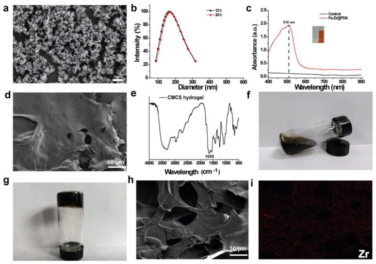
In recent years, multifunctional hydrogel nanoplatforms for the synergistic treatment of tumors have received a great deal of attention. Here, we prepared an iron/zirconium/polydopamine/carboxymethyl chitosan hydrogel with Fenton and photothermal effects, promising for future use in the field of synergistic therapy and prevention of tumor recurrence. The iron (Fe)-zirconium (Zr)@ polydopamine (PDA) nanoparticles were synthesized by a simple one-pot hydrothermal method using iron (III) chloride hexahydrate (FeCl3•6H2O), zirconium tetrachloride (ZrCl4), and dopamine, followed by activation of the carboxyl group of carboxymethyl chitosan (CMCS) using 1-(3-Dimethylaminopropyl)-3-ethylcarbodiimide hydrochloride (EDC)/N(4)-hydroxycytidine (NHS). Finally, the Fe-Zr@PDA nanoparticles and the activated CMCS were mixed to form a hydrogel. On the one side, Fe ions can use hydrogen peroxide (H2O2) which is rich in the tumor microenvironment (TME) to produce toxic hydroxyl radicals (•OH) and kill tumor cells, and Zr can also enhance the Fenton effect; on the other side, the excellent photothermal conversion efficiency of the incorporated PDA is used to kill tumor cells under the irradiation of near-infrared light. The ability of Fe-Zr@PDA@CMCS hydrogel to produce •OH and the ability of photothermal conversion were verified in vitro, and swelling and degradation experiments confirmed the effective release and good degradation of this hydrogel in an acidic environment. The multifunctional hydrogel is biologically safe at both cellular and animal levels. Therefore, this hydrogel has a wide range of applications in the synergistic treatment of tumors and the prevention of recurrence.

Keywords: Fenton reaction; chemodynamic therapy; hydrogel; photothermal therapy.


Conflict of interest statement

The authors declare no conflict of interest.

Figures

Scheme 1
Scheme 1
A practical and working schematic of a multifunctional hydrogel that can be injected for future use in killing tumors and inhibiting their postoperative recurrence (CDT: chemodynamic therapies; PTT: photothermal therapy).
Figure 1
Figure 1
(a) SEM image of Fe–Zr@PDA nanoparticle; (b) the hydration kinetic diameter of Fe–Zr@PDA in H2O; (c) the light absorption of phenanthroline solution with and without co-incubation with Fe–Zr@PDA; (d) SEM image of CMCS hydrogel; (e) FTIR spectra of CMCS hydrogel; (f) flowing solution state of Fe–Zr@PDA@CMCS hydrogel; (g) solidified state of Fe–Zr@PDA@CMCS hydrogel; (h) SEM image of Fe–Zr@PDA@CMCS hydrogel; (i) elemental distribution of Zr in Fe–Zr@PDA@CMCS hydrogel.
Figure 2
Figure 2
(a) Temperature variation of Fe–Zr@PDA@CMCS hydrogel with different Fe–Zr@PDA doping levels under 808 nm laser irradiation; (b) infrared thermographs correspond to (a), colors indicate different temperatures; (c) the temperature profiles of Fe–Zr@PDA@CMCS hydrogel irradiated with NIR laser with different power densities of 0.5, 0.8, and 1.0 W/cm2; (d) infrared thermographs correspond to (c), colors indicate different temperatures; (e) results from photothermal cycling tests results of Fe–Zr@PDA@CMCS hydrogel after 8 irradiation and cooling cycles; (f) time constant curves for Fe–Zr@PDA@CMCS hydrogel (808 nm, 1.0 W/cm2); (g) in vitro η values for Fe–Zr@PDA@CMCS hydrogel.
Figure 3
Figure 3
(a) Experimental results and corresponding digital camera photos of the reaction of different concentrations of Fe–Zr@PDA nanoenzymes with TMB and H2O2 to produce •OH; (b) absorbance values of different concentrations of Fe–Zr@PDA@CMCS after co-incubation with TMB and H2O2, and digital photographs of the supernatant photographic images; (c) degradation rates of Fe–Zr@PDA@CMCS hydrogel in CBS and PBS solutions; (d) swelling rate of Fe–Zr@PDA@CMCS hydrogel in ultrapure water, CBS solution, and PBS solution.
Figure 4
Figure 4
(a) Cell viability of Fe–Zr@PDA@CMCS hydrogel extracts at 1 mg/mL, 2.5 mg/mL, and 5 mg/mL co-cultured with mouse fibroblasts (L929 cell line) for 24 h and 48 h; (b) AM-PI stained images of L929 cell lines co-cultured with Fe–Zr@PDA@CMCS hydrogel extracts at 1 mg/mL, 2.5 mg/mL, and 5 mg/mL for 24 h and 48 h; (c) hemolysis ratio of rat erythrocytes after co-culture with different concentrations of Fe–Zr@PDA@CMCS hydrogel; (d) photos of rat erythrocytes after centrifugation.
Figure 5
Figure 5
(a) Changes in body weight of Fe–Zr@PDA@CMCS hydrogel treated mice and normal mice (control) at day 28; (b) results of serum biochemical parameters of Fe–Zr@PDA@CMCS hydrogel treated mice and normal mice (control); (c) H&E staining of the main organs of the Fe–Zr@PDA@CMCS hydrogel treated mice and normal mice (control). Bar = 200 μm.
Figure 6
Figure 6
Routine blood test data of Fe–Zr@PDA@CMCS hydrogel-treated mice and normal mice (control): (a) white blood cell (WBC), (b) red blood cell (RBC), (c) hemoglobin (HB), (d) red blood cell-specific volume (HCT), (e) mean corpuscular volume (MCV), (f) mean corpuscular hemoglobin (MCH), (g) mean corpuscular hemoglobin concentration (MCHC), (h) red cell volume distribution width (RDW), and (i) PLATELET (PLT).
Figure 7
Figure 7
(a) CCK-8 results of cells after different treatments; (bf) results of live and dead cell staining of SW1990 cells, morphological staining images of cells corresponding to CCK-8 results (b): L929 cells co-incubated with fresh medium; (c): L929 cells co-incubated with 100 μg/mL Fe–Zr@PDA nanoparticles; (d): L929 cells co-incubated with 100 μg/mL Fe–Zr@PDA@CMCS hydrogel extract; (e): L929 cells irradiated by 808 nm NIR, 1 W/cm2, 5 min pure photothermal irradiation; (f): fluorescent staining images of L929 cells after treatment with 100 μg/ mL Fe–Zr@PDA@CMCS hydrogel extract and 808 nm NIR, 1 W/cm2, by 5 min of light. Bar = 100 μm, **** p < 0.0001.

References

    1. Dolgin E. Cancer’s new normal. Nat. Cancer. 2021;2:1248–1250. doi: 10.1038/s43018-021-00304-7. - DOI - PubMed
    1. Wyld L., Audisio R.A., Poston G.J. The evolution of cancer surgery and future perspectives. Nat. Rev. Clin. Oncol. 2015;12:115–124. doi: 10.1038/nrclinonc.2014.191. - DOI - PubMed
    1. Frei E., III Curative cancer chemotherapy. Cancer Res. 1985;45:6523–6537. - PubMed
    1. Allen C., Her S., Jaffray D.A. Radiotherapy for Cancer: Present and Future. Adv. Drug Deliv. Rev. 2017;109:1–2. doi: 10.1016/j.addr.2017.01.004. - DOI - PubMed
    1. Jarosz-Biej M., Smolarczyk R., Cichoń T., Kułach N. Tumor Microenvironment as A “Game Changer” in Cancer Radiotherapy. Int. J. Mol. Sci. 2019;20:3212. doi: 10.3390/ijms20133212. - DOI - PMC - PubMed

LinkOut - more resources