. 2023 Jun 8;14(6):318.
doi: 10.3390/jfb14060318.

Decellularized Porcine Conjunctiva in Treating Severe Symblepharon

Affiliations

Decellularized Porcine Conjunctiva in Treating Severe Symblepharon

Fengmei Shan et al. J Funct Biomater. .

Abstract

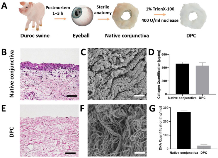
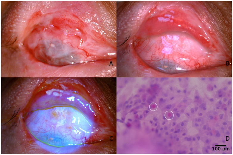
This prospective study aimed to evaluate the effectiveness of decellularized porcine conjunctiva (DPC) in the management of severe symblepharon. Sixteen patients with severe symblepharon were enrolled in this study. After symblepharon lysis and Mitomycin C (MMC) application, tarsus defects were covered with residual autologous conjunctiva (AC), autologous oral mucosa (AOM), or DPC throughout the fornix, and DPC was used for all the exposed sclera. The outcomes were classified as complete success, partial success, or failure. Six symblepharon patients had chemical burns and ten had thermal burns. Tarsus defects were covered with DPC, AC, and AOM in two, three, and eleven cases, respectively. After an average follow-up of 20.0 ± 6 months, the anatomical outcomes observed were complete successes in twelve (three with AC+DPC, four with AC+AOM+DPC, and five with AOM+DPC) (75%) cases, partial successes in three (one with AOM+DPC and two with DPC+DPC) (18.75%) cases, and failure in one (with AOM+DPC) (6.25%) case. Before surgery, the depth of the narrowest part of the conjunctival sac was 0.59 ± 0.76 mm (range, 0-2 mm), tear fluid quantity (Schirmer II tests) was 12.5 ± 2.26 mm (range, 10-16 mm), and the distance of the eye rotation toward the opposite direction of the symblepharon was 3.75 ± 1.39 mm (range, 2-7 mm). The fornix depths increased to 7.53 ± 1.64 mm (range, 3-9 mm), eye movement was significantly improved, and the distance of eye movement reaching 6.56 ± 1.24 mm (range, 4-8 mm) 1 month after the operation; the postoperative Schirmer II test (12.06 ± 2.90 mm, range, 6-17 mm) was similar to that before surgery. Goblet cells were finally found in fifteen patients by conjunctival impression cytology in the transplantation area of DPC, except for one patient who failed. DPC could be considered an alternative for ocular surface reconstruction of severe symblepharon. Covering tarsal defects with autologous mucosa is necessary for extensive reconstruction of the ocular surface.

Keywords: conjunctival reconstruction; decellularized extracellular matrix; oral mucosal transplantation; symblepharon.


Conflict of interest statement

The authors declare no conflict of interest.

Figures

Figure 1
Figure 1
Duroc swine without virus infection and medical history were selected. The conjunctivas were isolated from porcine eyes aseptically within 1–3 h postmortem. The native conjunctivas without epithelium were incubated in super nuclease with 400 U/mL and 1% TrionX-100 at 37 °C for 2 h. After repeated washing, the prepared DPCs were sterilized and stored at −20 °C (A). Histological comparison of native porcine conjunctiva (NPC) (B) and decellularized conjunctiva (E) by H&E staining. Scale bar, 50 μm. Scanning electron microscope image of NPC (C) and DPC (F), scale bar, 10 μm. Comparison of collagen content between NPC and DPC (D), comparison of DNA content between NPC and DPC (G).
Figure 2
Figure 2
Schematic graphic of three different kinds of surgical strategies during reconstruction of conjunctival fornix. The conjunctival defect was mended with DPC (blue) from the corneal limbus to the deep fornix.The palpebral conjunctival defect was mended with residual conjunctiva (pink) (A), residual conjunctiva and AOM (yellow) (B) or totally AOM (C). Transcutaneous double-armed 1–0 silk threads were used to fixed graft deep into the fornix (red).
Figure 3
Figure 3
Photos showing severe symblepharon before and after fonix reconstruction with DPC. Case 7 with grade Ⅲ symblepharon in the inferior fornix (A) and complete success achieved with deep fornix one month after operation (B). Case 9 with grade IV symblepharon in the supranasal fornix (C) and complete success achieved with deep fornix 12 months after operaton (D). Case 12 with grade IV symblepharon in the inferior fornix with pseudopterygium and upgaze restriction (E), and a deep fornix without inflammation and upgaze restriction was observed at 11-month after operation (F). Case 2 with extensive grade IV symblepharon in the inferior fornix (G), and partial success was achieved with focal recurrence of symblepharon 6-month after operation (H).
Figure 4
Figure 4
Photos of case 6 before and after fornix reconstruction. The upper conjunctival sac exhibited third-degree C symblepharon, accompanied by slightly hyperemic, pseudopterygium and restricted downward gaze (A). One month after operation, the conjunctival sac was significantly deepened and the residual autologous conjunctiva, AOM, and DPC all survived. The hyperemia of the ocular surface was reduced and down gaze restriction was released (B). Fluorescein staining shows complete epithelization of DPC graft (C). Goblet cells were found using impression cytology 1 month after surgery (the white circle) (D).

Similar articles

Cited by

References

    1. Sant’ Anna A.E., Hazarbassanov R.M., de Freitas D., Gomes J.Á. Minor salivary glands and labial mucous membrane graft in the treatment of severe symblepharon and dry eye in patients with Stevens-Johnson syndrome. Br. J. Ophthalmol. 2012;96:234–239. doi: 10.1136/bjo.2010.199901. - DOI - PubMed
    1. Senger B., Memar S.A., Ahmann A., Houser J.J., Doughty-McDonald L. Dermatologic and ophthalmologic treatment of erythema multiforme major: A case report. Cureus. 2021;13:e20854. doi: 10.7759/cureus.20854. - DOI - PMC - PubMed
    1. Aslan Katırcıoğlu Y., Kaderli A., Şingar Özdemir E., Örnek F. Clinical Results of the Use of Amniotic Membrane Transplantation Alone or in Combination with Adjuvant Therapies in Conjunctival Fornix Reconstruction. Turk. J. Ophthalmol. 2022;52:237–245. doi: 10.4274/tjo.galenos.2021.77019. - DOI - PMC - PubMed
    1. Kheirkhah A., Ghaffari R., Kaghazkanani R., Hashemi H., Behrouz M.J., Raju V.K. A combined approach of amniotic membrane and oral mucosa transplantation for fornix reconstruction in severe symblepharon. Cornea. 2013;32:155–160. doi: 10.1097/ICO.0b013e318247983d. - DOI - PubMed
    1. Kuckelkorn R., Schrage N., Redbrake C., Kottek A., Reim M. Autologous transplantation of nasal mucosa after severe chemical and thermal eye burns. Acta Ophthalmol. Scand. 1996;74:442–448. doi: 10.1111/j.1600-0420.1996.tb00596.x. - DOI - PubMed

LinkOut - more resources