. 2023 Jun 15;14(6):322.
doi: 10.3390/jfb14060322.

PEGylated Paclitaxel Nanomedicine Meets 3D Confinement: Cytotoxicity and Cell Behaviors

Affiliations

PEGylated Paclitaxel Nanomedicine Meets 3D Confinement: Cytotoxicity and Cell Behaviors

Wenhai Lin et al. J Funct Biomater. .

Abstract

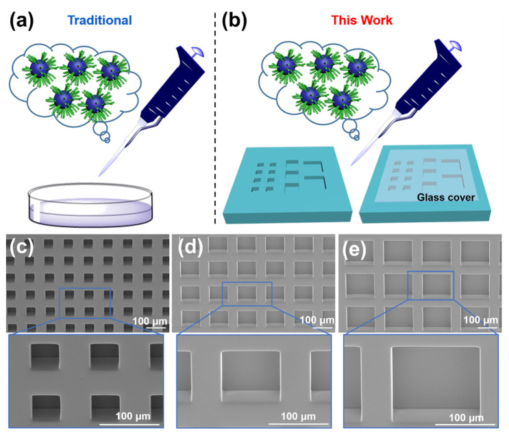
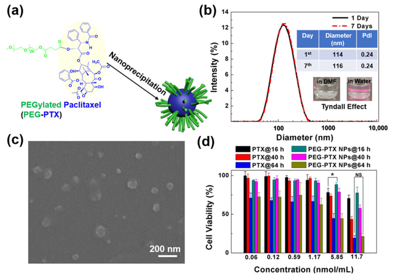
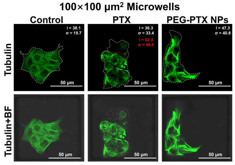
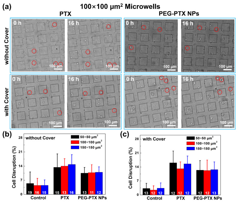
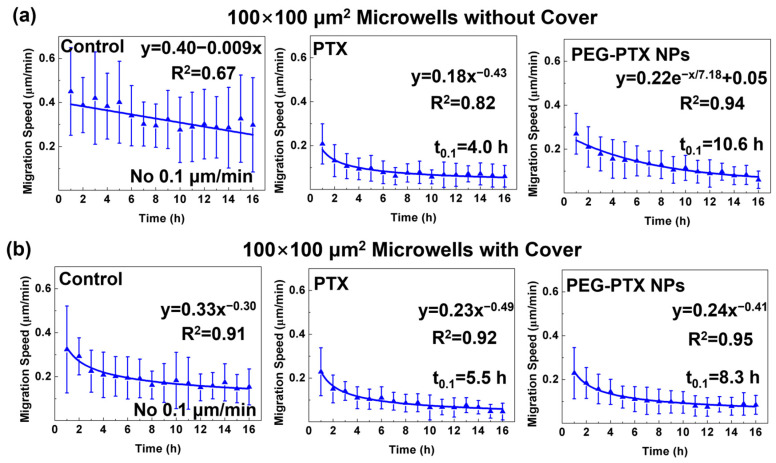
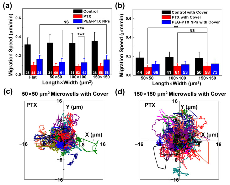
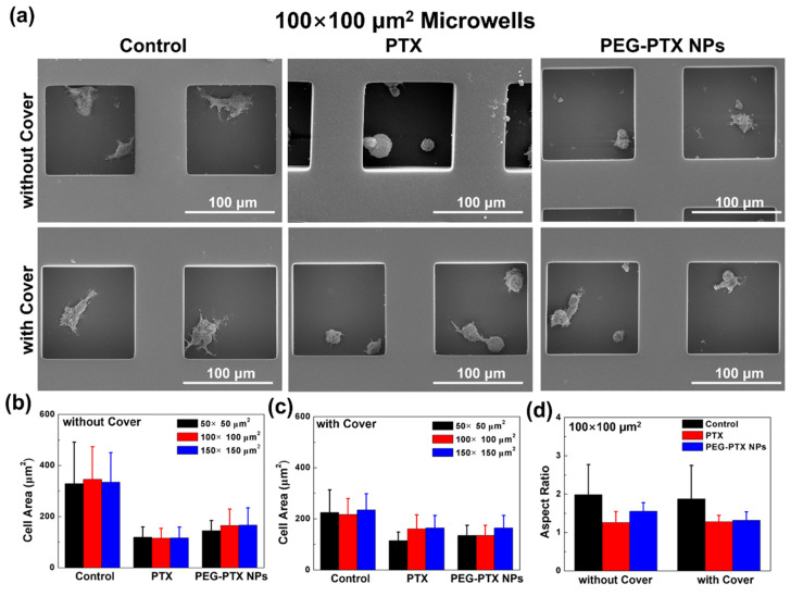
Investigating the effect of nanomedicines on cancer cell behavior in three-dimensional (3D) platforms is beneficial for evaluating and developing novel antitumor nanomedicines in vitro. While the cytotoxicity of nanomedicines on cancer cells has been widely studied on two-dimensional flat surfaces, there is little work using 3D confinement to assess their effects. This study aims to address this gap by applying PEGylated paclitaxel nanoparticles (PEG-PTX NPs) for the first time to treat nasopharyngeal carcinoma (NPC43) cells in 3D confinement consisting of microwells with different sizes and a glass cover. The cytotoxicity of the small molecule drug paclitaxel (PTX) and PEG-PTX NPs was studied in microwells with sizes of 50 × 50, 100 × 100, and 150 × 150 μm2 both with and without a concealed top cover. The impact of microwell confinement with varying sizes and concealment on the cytotoxicity of PTX and PEG-PTX NPs was analyzed by assessing NPC43 cell viability, migration speed, and cell morphology following treatment. Overall, microwell isolation was found to suppress drug cytotoxicity, and differences were observed in the time-dependent effects of PTX and PEG-PTX NPs on NPC43 cells in isolated and concealed microenvironments. These results not only demonstrate the effect of 3D confinement on nanomedicine cytotoxicity and cell behaviors but also provide a novel method to screen anticancer drugs and evaluate cell behaviors in vitro.

Keywords: 3D confinement; cell migration; microwell; nanomedicine; paclitaxel.


Conflict of interest statement

The authors declare no conflict of interest.

Figures

Figure 1
Figure 1
Schematic illustration to study effect of nanomedicines on cell behaviors and cytotoxicity of PEG-PTX NPs on (a) flat surface and (b) 3D platforms of microwell arrays without and with glass cover. Green: PEG; Blue: PTX. Scanning electron micrographs of (c) 50 × 50 μm2, (d) 100 × 100 μm2, and (e) 150 × 150 μm2 polydimethylsiloxane microwells.
Figure 2
Figure 2
Characterization of PEGylated paclitaxel nanoparticles (PEG-PTX NPs). (a) Schematic illustration of PEG-PTX NPs synthesis. (b) Size distribution and 7-day stability of PEG-PTX NPs measured by dynamic light scattering. Inserted images: PEG-PTX dissolved in dimethylformamide (left) and PEG-PTX NPs in water (right). (c) Scanning electron micrographs of PEG-PTX NPs. (d) Cell viability of NPC43 cells treated with PTX and PEG-PTX NPs for 16, 40, and 64 h, respectively. One-way ANOVA and Tukey’s post-hoc tests, NS—not significant; * p < 0.05.
Figure 3
Figure 3
Confocal laser scanning micrographs of NPC43 cells incubated with PTX and PEG-PTX NPs for 16 h in 100 × 100 μm2 microwells. Tubulin in cells was stained by tubulin-tracker green (green fluorescence) and overlays of tubulin and bright field (BF) images. Average value (I) and standard deviation (σ) of fluorescent intensity were larger after drug treatment. Red dotted lines outlined the boundary between ruptured cells and intact cells. Statistics presented in red excluded the ruptured cells. Concentration of PTX and PEG-PTX NPs were both 5.85 nmol/mL.
Figure 4
Figure 4
Comparison of NPC43 cell disruption ratio with different treatments. (a) Optical images of NPC43 cells incubated with PTX and PEG-PTX NPs for 16 h in 100 × 100 μm2 microwells without and with glass cover. Cell disruption highlighted in red circles. Percentage of cell disruption after PTX and PEG-PTX NP addition for 16 h in different microwells (b) without cover and (c) with cover. Number of NPC43 cells counted marked in white. Concentration of PTX and PEG-PTX NPs were both 5.85 nmol/mL.
Figure 5
Figure 5
Trends of NPC43 cell migration speed in 100 × 100 μm2 microwells (a) without cover and (b) with cover after different treatments. These groups include control without any treatment, NPC43 cells treated with PTX, and NPC43 cells treated with PEG-PTX NPs. t0.1 represents time when migration speed was equal to 0.1 μm/min. Concentration of PTX and PEG-PTX NPs were both 5.85 nmol/mL.
Figure 6
Figure 6
NPC43 cell migration behavior in microwells without and with cover. (a) Migration speed of NPC43 cells with PTX and PEG-PTX NPs treatments over 16 h in microwells (a) without cover and (b) with cover. Cell migration trajectories of NPC43 cells over 16 h with PTX added in (c) 50 × 50 μm2 microwells with cover and (d) 150 × 150 μm2 microwells with cover. Starting points of cell migration trajectories are (0, 0), trajectories of individual cells labeled with different colors. One-way ANOVA and Tukey’s post-hoc tests, NS—not significant, ** p < 0.01, and *** p < 0.001. Number of NPC43 cells counted in white. Concentration of PTX and PEG-PTX NPs were both 5.85 nmol/mL.
Figure 7
Figure 7
NPC43 cell morphology after various treatments. (a) Scanning electron micrographs of NPC43 cells with PTX and PEG-PTX NPs added over 16 h in 100 × 100 μm2 microwells without and with cover. Cell area after cells treated with PTX and PEG-PTX NPs for 16 h in microwells (b) without cover and (c) with cover. (d) Cell aspect ratio after NPC43 cells treated with PTX and PEG-PTX NPs for 16 h in 100 × 100 μm2 microwells without and with cover. Number of NPC43 cells counted in white. Concentration of PTX and PEG-PTX NPs were both 5.85 nmol/mL.

Similar articles

Cited by

References

    1. Kargozar S., Mollazadeh S., Kermani F., Webster T.J., Nazarnezhad S., Hamzehlou S., Baino F. Hydroxyapatite nanoparticles for improved cancer theranostics. J. Funct. Biomater. 2022;13:100. doi: 10.3390/jfb13030100. - DOI - PMC - PubMed
    1. Crawley J.W., Gow I.E., Lawes N., Kowalec I., Kabalan L., Catlow C.R.A., Logsdail A.J., Taylor S.H., Dummer N.F., Hutchings G.J. Heterogeneous trimetallic nanoparticles as catalysts. Chem. Rev. 2022;122:6795–6849. doi: 10.1021/acs.chemrev.1c00493. - DOI - PMC - PubMed
    1. Lin W., Colombani-Garay D., Huang L., Duan C., Han G. Tailoring nanoparticles based on boron dipyrromethene for cancer imaging and therapy. Wiley Interdiscip. Rev. Nanomed. Nanobiotechnol. 2020;12:e1627. doi: 10.1002/wnan.1627. - DOI - PubMed
    1. Song D., Xu Q. Engineering a Nano/Biointerface for Cell and Organ-Selective Drug Delivery. Langmuir. 2022;38:9092–9098. doi: 10.1021/acs.langmuir.2c01609. - DOI - PubMed
    1. Calzoni E., Cesaretti A., Polchi A., Di Michele A., Tancini B., Emiliani C. Biocompatible polymer nanoparticles for drug delivery applications in cancer and neurodegenerative disorder therapies. J. Funct. Biomater. 2019;10:4. doi: 10.3390/jfb10010004. - DOI - PMC - PubMed

LinkOut - more resources