Review
. 2023 May 30;10(6):240.
doi: 10.3390/jcdd10060240.

Recasting Current Knowledge of Human Fetal Circulation: The Importance of Computational Models

Affiliations
Review

Recasting Current Knowledge of Human Fetal Circulation: The Importance of Computational Models

Daibo Zhang et al. J Cardiovasc Dev Dis. .

Abstract

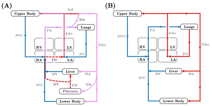
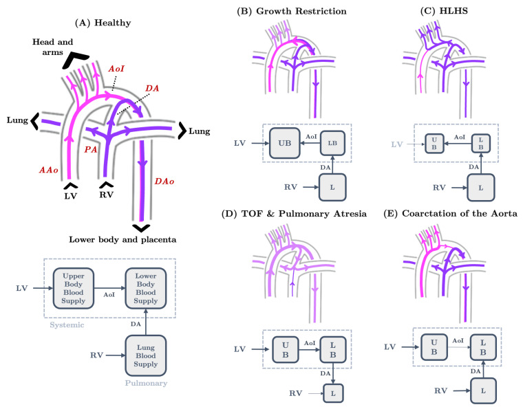
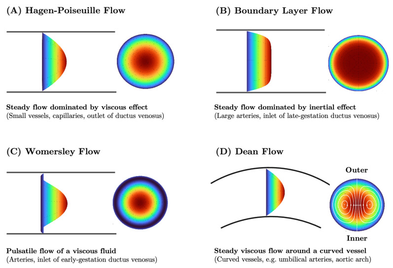
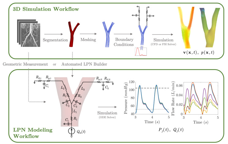
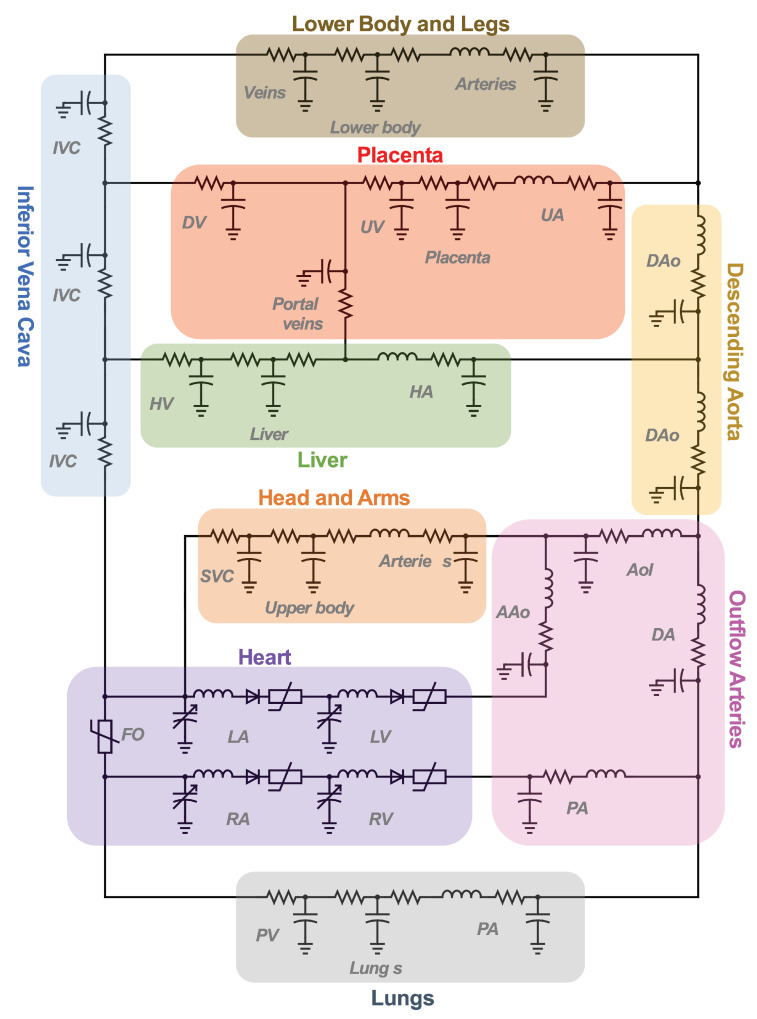
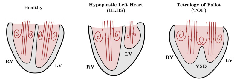
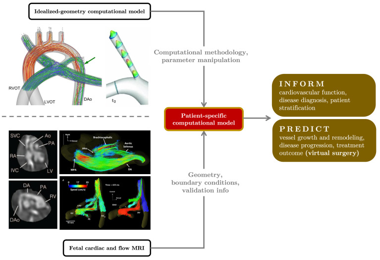
Computational hemodynamic simulations are becoming increasingly important for cardiovascular research and clinical practice, yet incorporating numerical simulations of human fetal circulation is relatively underutilized and underdeveloped. The fetus possesses unique vascular shunts to appropriately distribute oxygen and nutrients acquired from the placenta, adding complexity and adaptability to blood flow patterns within the fetal vascular network. Perturbations to fetal circulation compromise fetal growth and trigger the abnormal cardiovascular remodeling that underlies congenital heart defects. Computational modeling can be used to elucidate complex blood flow patterns in the fetal circulatory system for normal versus abnormal development. We present an overview of fetal cardiovascular physiology and its evolution from being investigated with invasive experiments and primitive imaging techniques to advanced imaging (4D MRI and ultrasound) and computational modeling. We introduce the theoretical backgrounds of both lumped-parameter networks and three-dimensional computational fluid dynamic simulations of the cardiovascular system. We subsequently summarize existing modeling studies of human fetal circulation along with their limitations and challenges. Finally, we highlight opportunities for improved fetal circulation models.

Keywords: cardiovascular lumped-parameter networks; computational fluid dynamics; congenital heart defects; fetal circulation; growth restriction; hemodynamics; patient-specific modeling; pediatric cardiology.


Conflict of interest statement

The authors declare no conflict of interest.

Figures

Figure 1
Figure 1
Blood flow in the fetal circulatory system (A) versus in the adult circulatory system (B). Dashed lines refer to shunts that are unique to fetal circulation. Arrows indicate flow direction. Line colors correspond to oxygenation levels with blue for oxygen-poor blood, red for oxygen-rich blood, and purple for mixed oxygenated and deoxygenated flow. Numerical values for oxygen saturation can be found in Appendix A. AAo—ascending aorta, DAo—descending aorta, PV—pulmonary vein, SVC—superior vena cava, PA—pulmonary artery, DA—ductus arteriosus, IVC—inferior vena vava, UA—umbilical arteries, DV—ductus venosus, UV—umbilical vein, RA/LA—right/left atrium, RV/LV—right/left ventricle, FO—foramen ovale.
Figure 2
Figure 2
Blood flow path in the human fetal great vessel network as produced by 3D simulations. (A) Under healthy conditions, blood flow from the PA to the DAo through the DA and from the AAo to the DAo through the AoI. (B) In growth-restricted fetuses, AoI blood flow reverses due to increases in upper body blood supply caused by decreases in brain vascular resistance. (C) In HLHS fetuses, RV flow also supplies the upper body systemic circulation due to an underdeveloped LV, causing flow reversal in the AoI. (D) In TOF or pulmonary artresia, systemic circulation rescues the inadequate pulmonary blood supply, causing flow reversal in the DA. (E) With coarctation of the aorta, flow reveresal is not observed in the AoI or the DA; rather, AoI flow is greatly reduced due to aortic stenosis. Pathline color correlates with oxygen saturation with pink indicating a higher oxygenation level than purple. Note that left ventricular output is more oxygen-rich than right ventricular output (Appendix A). AAo—ascending aorta, AoI—aortic isthmus, DA—ductus arteriosus, DAo—descending aorta, HLHS—hypoplastic left heart syndrome, LV—left ventricle, PA—pulmonary arteries, RV—right ventricle, TOF—tetralogy of Fallot.
Figure 3
Figure 3
The anatomy and functional properties of the umbilical circulation network. The portion of umbilical arteries and veins outside of the fetal body is contained in the umbilical cord. The coiled geometry of the umbilical vessels maintains a stable mechanical stress environment as the cord contorts due to fetal motion and may contribute to fetal thermal regulation. When the cord is overcoiled (characterized by an elevated umbilical coiling index), vascular resistance increases, potentially implicating cord overcoiling in growth restriction. When the coil diameter reduces, umbilical cord stricture occurs and wall shear stress in the vessels increases, which may lead to stenosis and the formation of thrombosis. Line colors indicate highly oxygenated blood (red) and moderately oxygenated blood (pink) (Appendix A for values). LV—left ventricle, IVC—inferior vena cava, DV—ductus venosus, DAo—descending aorta, CIA—common iliac artery, IIA—internal iliac artery, UA—umbilical artery, WSS—wall shear stress.
Figure 4
Figure 4
Hemodynamic flow profiles experienced in various cardiovascular simulations (A) Poiseuille flow is dominated by viscous effects and marked by a parabolic profile. Commonly seen in small vessels, capillaries, outlet of ductus venosus. (B) Boundary layer flow is dominated by inertial effect and marked by a rectangular, plug-like, profile. Often appears in large arteries, inlet of late-gestation ductus venosus. (C) Womersley flow exhibits a small amount of flow reversal near the vessel wall due to competing viscous and inertial forces. Typical of arteries and early gestation ductus venosus inlet flow. (D) Dean flow in minimally curved vessels produces a parabolic primary flow pattern as viscosity is the dominant phenomenon. A pair of counter-rotating vortices constitutes the secondary flow due to the combined effect of inertial and centripetal forces. Plots created via [101].
Figure 5
Figure 5
Computational workflow for patient-specific 3D or lumped parameter network (LPN) hemodynamic simulation. For 3D simulations, the steps include segmentation, meshing, defining of boundary conditions, and simulation. A time-varying flow curve is imposed at the inlet and a RCR Windkessel model representing downstream vasculature at the outlet. For LPN simulations, the Windkessel model can be constructed from geometric measurements or automatically from 3D vessel models. Large vessel segments are represented by RLC circuits, a small peripheral vascular network is represented by RCR circuits, and an inflow curve is represented as a current source.
Figure 6
Figure 6
Representative fetal LPN circuit. Note how it relies heavily on RLC components as a basic vessel building block.
Figure 7
Figure 7
Schematic of diastolic blood flow and vortex patterns in fetal ventricles. In the healthy fetal heart, diastolic vortex rings are attached to the ventricular walls, generating high WSS. With hypoplastic left heart syndrome (HLHS), the diastolic vortex ring in the underdeveloped left ventricle does not contact the ventricular wall, so left ventricle WSS is persistently low. With tetralogy of Fallot (TOF), diastolic vortex rings in the two ventricles interact due to flow across the ventricular septal defect (VSD), applying high WSS on the ventricular septum. LV—left ventricle, RV—right ventricle.
Figure 8
Figure 8
Outlook for patient-specific fetal hemodynamic modeling. Vascular morphology and flow information provided by fetal MRI ([84,183]) can be integrated with existing computational frameworks established using idealized geometry models (examples from [27,170]) to create patient-specific models of fetal circulation. The detailed blood flow information and predicative capability provided by computational modeling provide patient-specific insights that can drive physiology studies, disease characterizations, and clinical decision making.

Similar articles

Cited by

References

    1. Rana M.S., Sizarov A., Christoffels V.M., Moorman A.F. Development of the human aortic arch system captured in an interactive three-dimensional reference model. Am. J. Med. Genet. Part A. 2014;164:1372–1383. doi: 10.1002/ajmg.a.35881. - DOI - PubMed
    1. Yagel S., Kivilevitch Z., Cohen S.M., Valsky D.V., Messing B., Shen O., Achiron R. The fetal venous system, Part I: Normal embryology, anatomy, hemodynamics, ultrasound evaluation and Doppler investigation. Ultrasound Obstet. Gynecol. 2010;35:741–750. doi: 10.1002/uog.7618. - DOI - PubMed
    1. Yee K., Lui F. StatPearls [Internet] StatPearls Publishing; Treasure Island, FL, USA: 2021. Anatomy, Thorax, Heart Foramen Ovale. - PubMed
    1. Lindsey S.E., Butcher J.T., Yalcin H.C. Mechanical regulation of cardiac development. Front. Physiol. 2014;5:318. doi: 10.3389/fphys.2014.00318. - DOI - PMC - PubMed
    1. Chew L.C., Verma R.P. Fetal Growth Restriction. StatPearls Publishing; Treasure Island, FL, USA: 2021. - PubMed

LinkOut - more resources