. 2023 May 24;11(2):22.
doi: 10.3390/jdb11020022.

Osteoderm Development during the Regeneration Process in Eurylepis taeniolata Blyth, 1854 (Scincidae, Sauria, Squamata)

Affiliations

Osteoderm Development during the Regeneration Process in Eurylepis taeniolata Blyth, 1854 (Scincidae, Sauria, Squamata)

Gennady O Cherepanov et al. J Dev Biol. .

Abstract

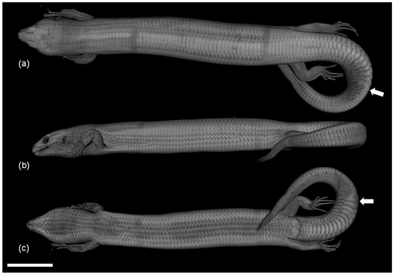
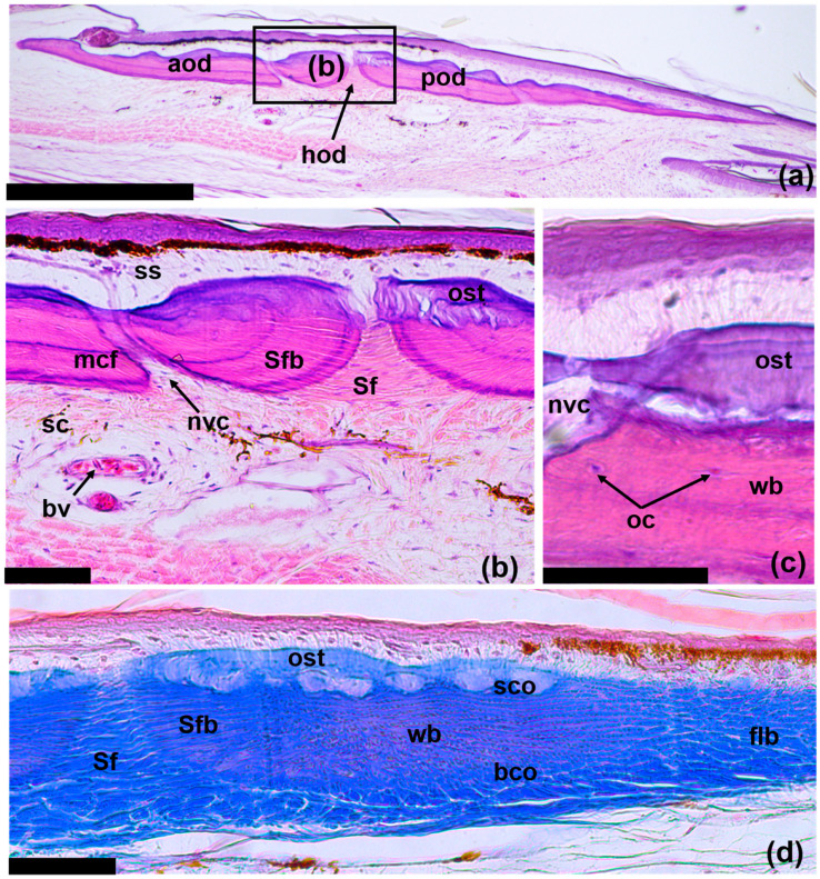
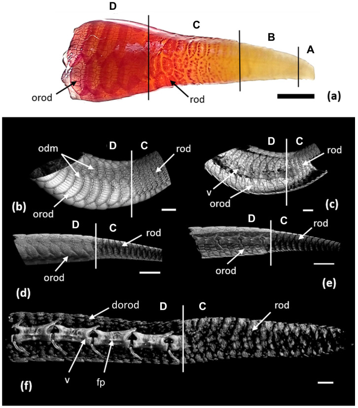
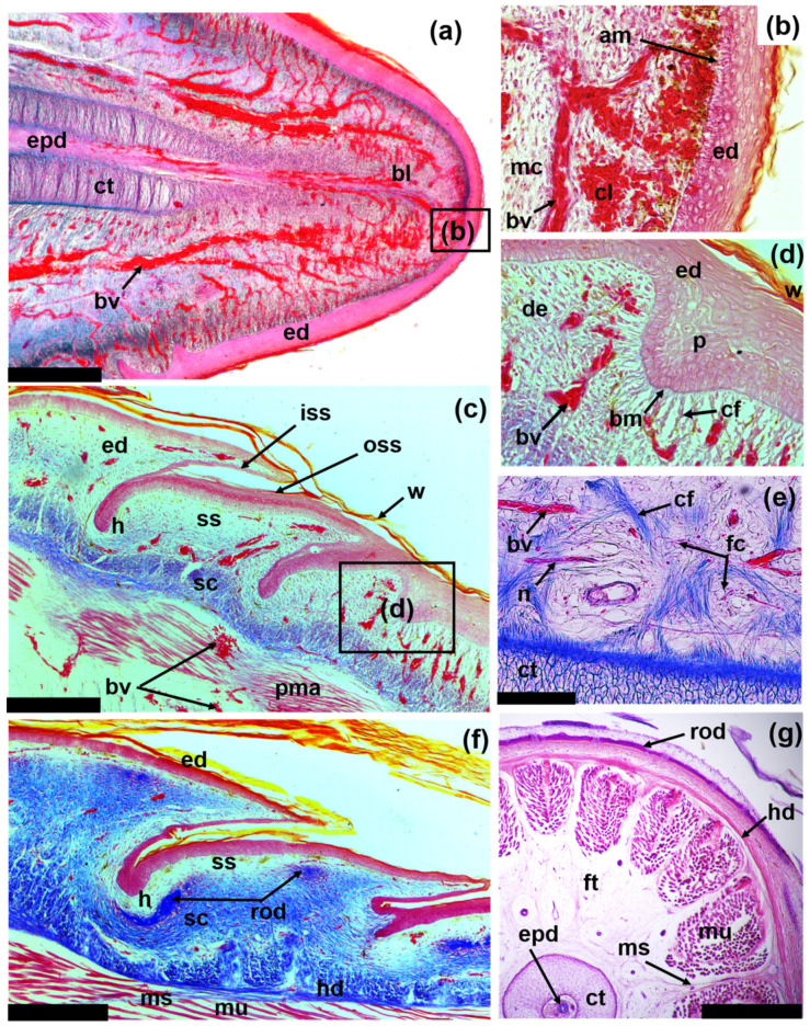
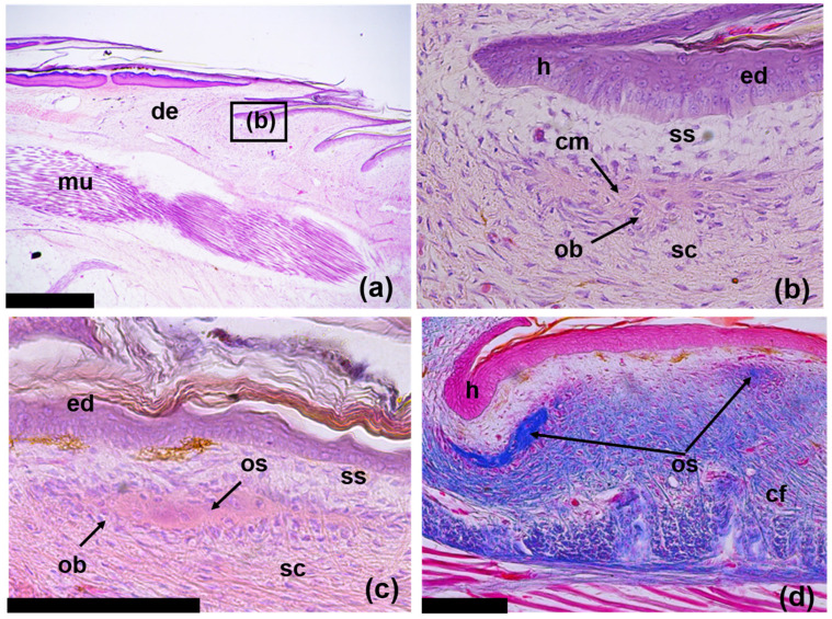
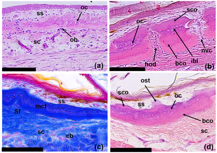
Osteoderms are bony structures that develop within the dermal layer of the skin in vertebrates and are very often found in different lizard families. Lizard osteoderms are diverse in topography, morphology, and microstructure. Of particular interest are the compound osteoderms of skinks, which are a complex of several bone elements known as osteodermites. We present new data on the development and regeneration of compound osteoderms based on the results of a histological and Computed Microtomography (micro-CT) study of a scincid lizard: Eurylepis taeniolata. The specimens studied are stored in the herpetological collections of the Saint-Petersburg State University and Zoological Institute of the Russian Academy of Sciences located in St. Petersburg, Russia. The topography of osteoderms in the integuments of the original tail area and its regenerated part was studied. A comparative histological description of the original and regenerated osteoderms of Eurylepis taeniolata is presented for the first time. The first description of the development of compound osteoderm microstructure in the process of caudal regeneration is also presented.

Keywords: Eurylepis taeniolata; anatomy; development; histology; osteoderms; regeneration.


Conflict of interest statement

The authors declare no conflict of interest. The funders had no role in the design of the study; in the collection, analyses, or interpretation of data; in the writing of the manuscript; or in the decision to publish the results.

Figures

Figure 1
Figure 1
Diagram of relationship between size (mm) and age (month) of regenerating tails in E. taeniolata. First number indicates number of individuals, in parentheses—number of generation (regenerate of first or second order).
Figure 2
Figure 2
Spatial organization of osteodermal cover of E. taeniolatus (R-5328-2) according to results of three-dimensional micro-CT reconstruction: (a) dorsal view; (b) lateral view; (c) ventral view. White arrows show area of autotomy. Scale bar: 20 mm.
Figure 3
Figure 3
Original tail osteoderms of E. taeniolata (ZISP # 5328) based on 3D micro-CT reconstruction: (a) outer and (b) inner surfaces. Abbreviations: aod, anterior osteodermites; iod, intermediate osteodermites; nvc, neurovascular canals; pod, posterior osteodermites. Scale bars: 2.5 mm.
Figure 4
Figure 4
Longitudinal sections of original tail osteoderms of E. taeniolata: histological preparations (a,b) SPbU # 1773; (c) SPbU # 1774; (d) SPbU # 1782. Abbreviations: aod, anterior osteodermite; bco, basal cortex; bv, blood vessel; flb, fibrolamellar (parallel-fibered) bone; hod, hinge region between osteodermites; nvc, neurovascular canal; oc, osteocytes; ost, osteodermine; pod, posterior osteodermite; sc, stratum compactum; sco, superficial cortex; Sf, Sharpey’s fibers; Sfb, Sharpey-fiber bone; ss, stratum superficiale; wb, woven bone. Sections are stained with Delafield’s hematoxylin, eosine (ac) and with Azan, azo-carmine B (d). Scale bars: (a) = 500 µm, (bd) = 50 µm.
Figure 5
Figure 5
Spatial organization of osteodermal integument of tail of E. taeniolata: (a) alizarin preparation (SPbU # R-125) with a regenerate 17.5 mm long (9.5-month-old); (bf) 3D micro-CT reconstruction of tail with regenerate: (b) ventral side and section in frontal plane; (c) tail of an adult specimen (ZISP # 5328); (d) left side and section in sagittal plane (e) of the tail of a subadult specimen (ZISP# 5327); (f) Left side of tail of a juvenile specimen (ZISP#5334). Abbreviations: dorod, developing original osteoderm; fp, fracture plane; odm, osteoderms; orod, original osteodermites; rod, regenerating osteodermites; v, vertebra. Vertical lines show key areas: A, zone of undifferentiated integument; B, zone of scale regeneration; C, zone of osteoderm regeneration; D, site of the original tail with developed osteodermal cover. Scale bars: (a) = 3.5 mm, (be) = 2.5 mm, (f) = 500 µm.
Figure 6
Figure 6
Frontal (af) and transverse (g) sections of regenerating tails of E. taeniolata: (ad,f) regenerate 5.5 mm long (SPbU # 1775); (e) regenerate 37 mm long (SPbU # 1782); (g) regenerate 49 mm long (SPbU # 1780). Abbreviations: am, aggregates of melanocytes; bl, blastema; bm, basal membrane; bv, blood vessel; cf, collagen fibers; cl, clot; ct, cartilaginous tube; de, dermis; ed, epidermis; epd, ependyma; fc, fat cells; ft, fat tissue; h, hinge region between scales; hd, hypodermis; iss, inner scale surface; mc, mesenchymal cells; ms, myosepta; mu, muscles; n, nerve; oss, outer scale surface; p, peg; pma, promuscle aggregates; rod, regenerating osteoderm; sc, stratum compactum; ss, stratum superficiale; w, corneous layer of the wound epidermis. Sections are stained with Azan, azo-carmine B (af) and with Delafield’s hematoxylin, eosine (g). Scale bar: (a,g) = 500 µm, (c,f) = 250 µm, (e) = 100 µm. Internal structures of regenerating tail extend through final stage of development (g). Cartilaginous tube is composed of hypertrophied cells, which are covered inside and outside with perichondrium. Canal of cartilaginous tube contains an ependyma and several large blood vessels. Tube is surrounded by a thick layer of adipose tissue (e) and contains main blood vessels and spinal nerves. Muscles are well differentiated. Myomeres are separated by longitudinal and transverse myosepta composed of connective tissue fibers.
Figure 7
Figure 7
Longitudinal skin sections of regenerating tails of E. taeniolata in area of developing osteoderms: (a,b) regenerate 5 mm long (SPbU # 1773); (c) regenerate 7 mm long (SPbU # 1776); (d) regenerate 5.5 mm long (SPbU # 1775). Abbreviations: cf, collagen fibers; cm, collagen matrix; de, dermis; ed, epidermis; h, hinge region between scales; mu, muscles; ob, osteoblasts; os, osteoid; sc, stratum compactum; ss, stratum superficiale. Sections are stained with Delafield’s hematoxylin, eosine (ac) and with Azan, azo-carmine B (d). Scale bars: (a) = 500 µm, (c,d) = 100 µm.
Figure 8
Figure 8
Transverse (a) and longitudinal (bd) skin sections of regenerating tails of E. taeniolata in area of developing osteoderms: (a,b) regenerate 27 mm long (SPbU # 1779); (c,d) regenerate 37 mm long (SPbU # 1782). Abbreviations: bco, basal cortex; cb, collagen bundles; hod, hinge region between osteodrmites; ibl, intermediate bone layer; mcf, mineralized collagen fibers; nvc, neurovascular canal; ob, osteoblasts; oc, osteocytes; ost, osteodermine; sc, stratum compactum; sco, superficial cortex; Sf, Sharpey’s fibers; ss, stratum superficiale. Sections are stained with Delafield’s hematoxylin, eosine (a,b,d) and with Azan, azo-carmine B (c). Scale bars: 100 µm.

Similar articles

Cited by

References

    1. Paluh D.J., Griffing A.H., Bauer A.M. Sheddable Armour: Identification of Osteoderms in the Integument of Geckolepis maculata (Gekkota) Afr. J. Herpetol. 2017;66:12–24. doi: 10.1080/21564574.2017.1281172. - DOI
    1. Marghoub A., Williams C.J.A., Leite J.V., Kirby A.C., Kéver L., Porro L.B., Barrett P.M., Bertazzo S., Abzhanov A., Vickaryous M., et al. Unravelling the Structural Variation of Lizard Osteoderms. Acta Biomater. 2022;146:306–316. doi: 10.1016/j.actbio.2022.05.004. - DOI - PubMed
    1. Vickaryous M., Williams C., Willan G., Kirby A., Herrel A., Kever L., Moazen M., Marghoub A., Rai S., Abzhanov A., et al. Histological Diversity and Evolution of Lizard Osteoderms. FASEB J. 2022;36 doi: 10.1096/fasebj.2022.36.S1.R3199. - DOI
    1. Moss M.L. Comparative Histology of Dermal Sclerifications in Reptiles. Acta Anat. 1969;73:510–533. doi: 10.1159/000143315. - DOI - PubMed
    1. Bochaton C., De Buffrenil V., Lemoine M., Bailon S., Ineich I. Body Location and Tail Regeneration Effects on Osteoderms Morphology—Are They Useful Tools for Systematic, Paleontology, and Skeletochronology in Diploglossine Lizards (Squamata, Anguidae)? J. Morphol. 2015;276:1333–1344. doi: 10.1002/jmor.20421. - DOI - PubMed

LinkOut - more resources