. 2023 May 31;11(2):24.
doi: 10.3390/jdb11020024.

Jak2 and Jaw Muscles Are Required for Buccopharyngeal Membrane Perforation during Mouth Development

Affiliations

Jak2 and Jaw Muscles Are Required for Buccopharyngeal Membrane Perforation during Mouth Development

Amanda J G Dickinson. J Dev Biol. .

Abstract

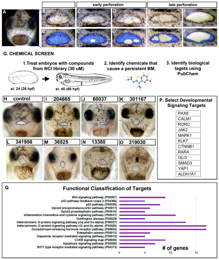
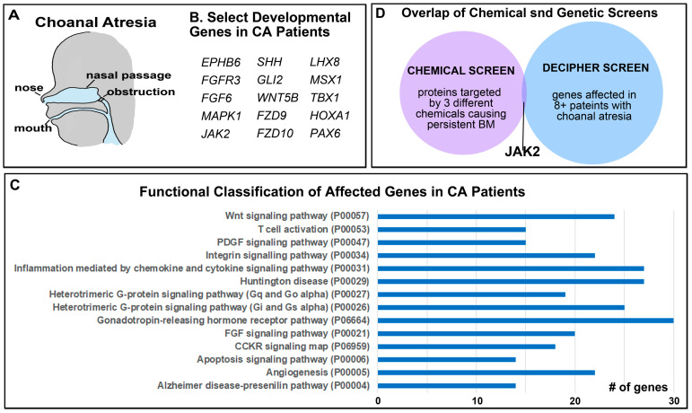
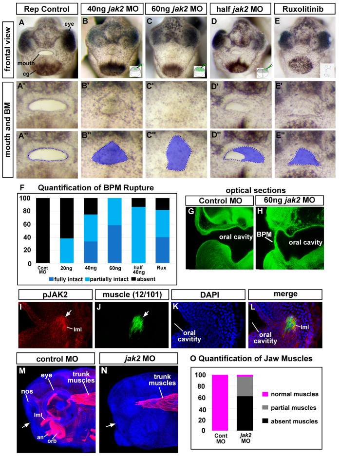
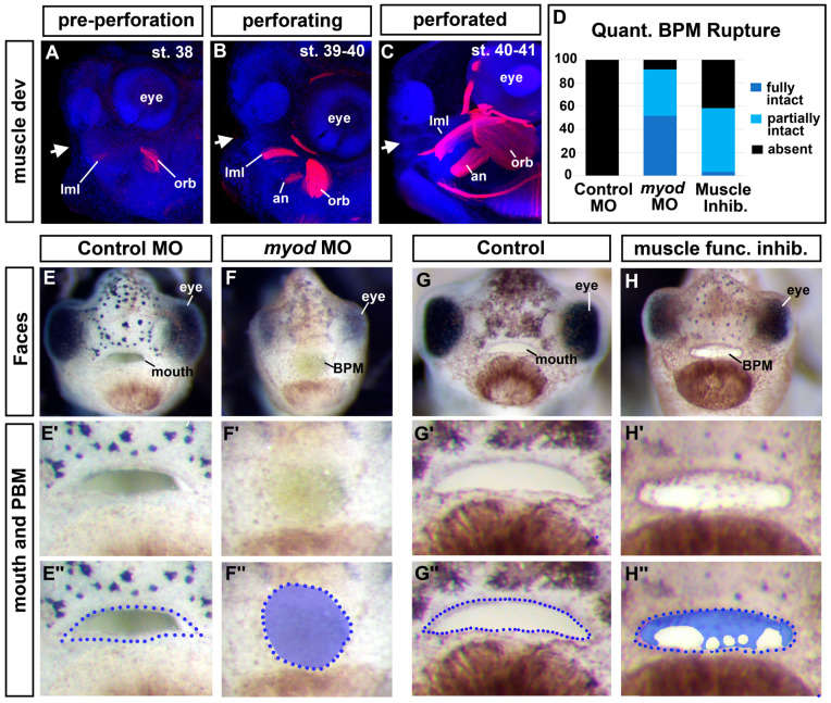
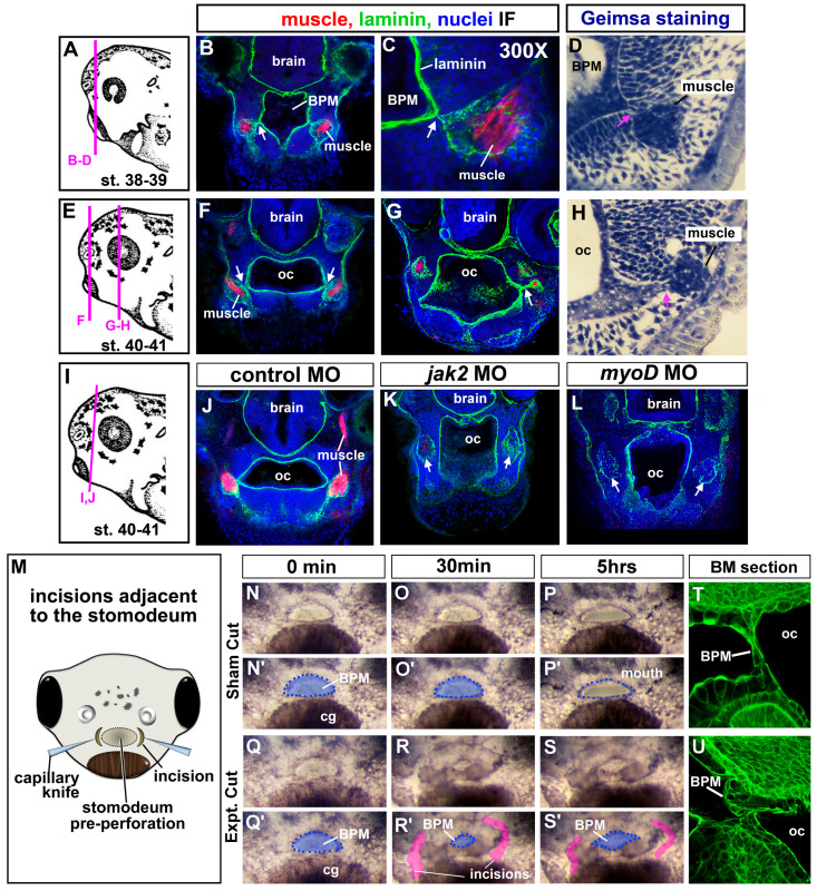
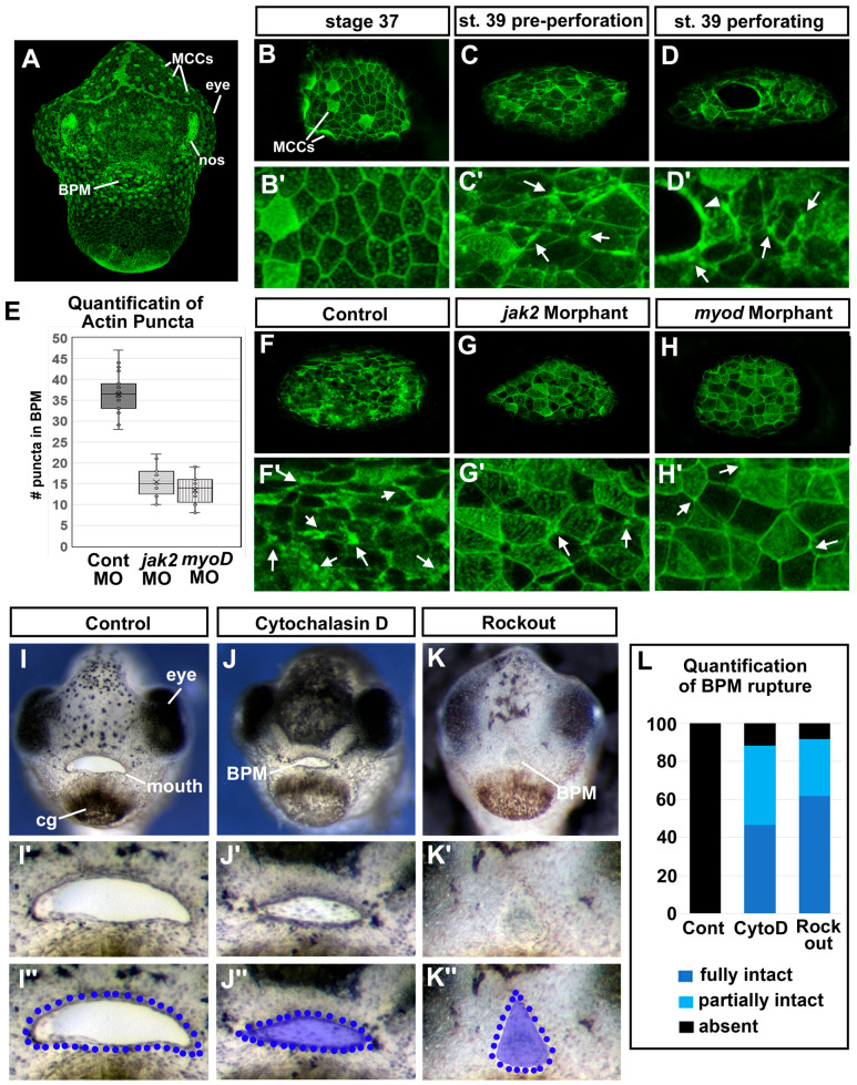
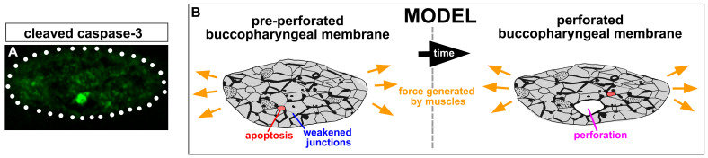
The mouth is a central feature of our face, without which we could not eat, breathe, or communicate. A critical and early event in mouth formation is the creation of a "hole" which connects the digestive system and the external environment. This hole, which has also been called the primary or embryonic mouth in vertebrates, is initially covered by a 1-2 cell layer thick structure called the buccopharyngeal membrane. When the buccopharyngeal membrane does not rupture, it impairs early mouth functions and may also lead to further craniofacial malformations. Using a chemical screen in an animal model (Xenopus laevis) and genetic data from humans, we determined that Janus kinase 2 (Jak2) has a role in buccopharyngeal membrane rupture. We have determined that decreased Jak2 function, using antisense morpholinos or a pharmacological antagonist, caused a persistent buccopharyngeal membrane as well as the loss of jaw muscles. Surprisingly, we observed that the jaw muscle compartments were connected to the oral epithelium that is continuous with the buccopharyngeal membrane. Severing such connections resulted in buccopharyngeal membrane buckling and persistence. We also noted puncta accumulation of F-actin, an indicator of tension, in the buccopharyngeal membrane during perforation. Taken together, the data has led us to a hypothesis that muscles are required to exert tension across the buccopharyngeal membrane, and such tension is necessary for its perforation.

Keywords: Xenopus laevis; buccopharyngeal membrane; choanal atresia; mouth.


Conflict of interest statement

The authors declare no conflict of interest.

Figures

Figure 1
Figure 1
Buccopharyngeal membrane rupture and chemical screen. (A) Frontal view of the face of an embryo at stage 39 prior to the perforation of the buccopharyngeal membrane. (BF’) magnified images of the mouth at progressive points of buccopharyngeal membrane perforation. In the prime labeled images, the buccopharyngeal membrane is shaded blue. Each image is from a different embryo that represents the most common appearance at each stage (from over 200 embryos examined in 10 biological replicates). (G) Schematic outlining the chemical screen. (HO) A subset of representative embryos treated with select chemicals causing a persistent buccopharyngeal membrane but with a stomodeum present. The presumptive mouth is outlined in blue dots. The National Service Center Number identifier is shown above each embryo. (P) Protein symbols of select targets of chemicals causing a persistent buccopharyngeal membrane. (Q) The top functional categories identified from the targets of chemicals causing a persistent buccopharyngeal membrane. The GO identifier is in brackets. Abbreviations: # = number, st. = stage.
Figure 2
Figure 2
Copy Number Variants in patients with Choanal Atresia. (A) Schematic showing where the airway blockage can occur in Choanal Atresia. (B) Select genes with copy number deletions identified in patients with Choanal Atresia known to be important in development. (C) The top functional categories identified from copy number deletions in patients with Choanal Atresia. The GO identifier is in brackets. (D) Overlap in genes identified in the chemical and genetic screens identifies JAK2. Abbreviations: # = number.
Figure 3
Figure 3
Jak2 knockdown affects the buccopharyngeal membrane and cranial muscles. (AE’’) Frontal view of the face of representative embryos at stages 40–41 showing ruptured or persisting buccopharyngeal membranes (images from n = 60, 3 biological replicates for each treatment). The bottom right corner shows an image of the injection site/stage. Prime-labeled images show magnified images of the mouth and the double-prime-labeled images show the same images with the buccopharyngeal membrane shaded in blue. (F) Shows relative percentages of embryos with an intact, partially intact, or absent buccopharyngeal membrane (n = 60 for each group, 3 biological replicates). (G,H) Optical sagittal section through the head of representative controls and jak2 morphants. The buccopharyngeal membrane is absent in the control and is present but thin in the jak2 morphants (n = 12, 2 biological replicates). (IL) Representative thick agarose section through the face showing phosphor-‘JAK2 and 12/101 immunofluorescence. (I) phospho-Jak2 (red), (J) 12/101 = muscle-specific antibody (green), (K) DAPI (blue), (L) merge. (M,N) Lateral views of representative embryos showing 12/101 muscle labeling (red) in control and jak2 morphants and counterstained with DAPI (blue). White arrows point to the location of the mouth. (O) Quantification of the presence of cranial muscles in control and jak2 morphants (n = 60, 2 biological reps). Abbreviations; BPM = buccopharyngeal membrane, nos = nostril, MO = morpholino, levator mandibulae longus = lml, orbithyoidus = orb, angularis = an.
Figure 4
Figure 4
Cranial muscles are required for buccopharyngeal membrane rupture. (AC) Lateral views of embryos showing cranial muscles (red) and counterstained with DAPI (blue) (best images from 10 embryos imaged at each stage). White arrows show the location of the developing mouth. (D) Proportion of embryos that had a fully intact, partially intact, or absent buccopharyngeal membrane after myoD knockdown or treatment with a muscle inhibitor (BTS) compared to controls (n = 60, 3 biological replicates for each treatment). (EH’’) Frontal views of representative embryos injected with myoD morpholinos or treated with an inhibitor of muscle function (BTS). Prime-labeled images show magnified images of the mouth, and the double-prime-labeled images show the buccopharyngeal membrane shaded in blue. Abbreviations: levator mandibulae longus = lml, orbithyoidus = orb, angularis = an, BPM = buccopharyngeal membrane.
Figure 5
Figure 5
(AL) Muscle and oral cavity connections. (A,E,I) Schematics of the lateral views of embryos showing locations of the corresponding sections. (B,C,F,G,JL) Transverse sections labeled with antibodies to detect muscle-specific protein (12/101 in red), laminin (green), and counterstained with DAPI (blue). Images were chosen from representative images taken from 20 different embryos in 2 biological replicates. White arrows indicate laminin that bridges the oral cavity and muscle compartments. (D,H) Plastic section stained with Giemsa showing representative images of the association between muscle and oral cavities (based on sections of 20 embryos in 2 biological replicates). Pink arrows indicate the connections between the muscle and the oral cavity. (M) Schematic of a frontal view showing the location of the surgical incisions. (NS’) Shows the embryonic mouth and buccopharyngeal membrane in representative embryos before (0 min) and after sham or incisions (at 30 min and 5 h). Prime images show the buccopharyngeal membrane shaded in blue and the mouth outlined in blue dots. Representative images chosen from 50 embryos performed over 5 biological replicates. In R and S, the incised tissue is colored pink. (T,U) representative embryos 30 min after surgeries (or sham) sectioned and labeled with phalloidin to show the cellular arrangements in the buccopharyngeal membrane. Shows representative images taken from a total of 10 embryos in 2 biological replicates, Abbreviations: oc = oral cavity, BPM = buccopharyngeal membrane, cg = cement gland.
Figure 6
Figure 6
Actin dynamics and buccopharyngeal perforation. (AG) Actin dynamics revealed by phalloidin labeling during buccopharyngeal membrane perforation. (A) Shows a frontal view of the face at stage 39 (representative from 30 embryos in 4 biological replicates). (B) The buccopharyngeal membrane at stage 37 when the mouth shape is round. (C) The buccopharyngeal membrane at stage 39 just prior to perforation. Note the puncta spots and accumulation of F-actin (white arrows). (D) The buccopharyngeal membrane during perforation shows accumulation of F-actin surrounding the holes. (EH’) The buccopharyngeal membranes of control morphants (F), jak2 morphants (G), and myod morphants (H). (IK’’) Images of representative embryos after treatment with cytochalasin D (1 μM) and Rockout (100 μM) from stages 37–40. Quantification of buccopharyngeal membrane rupture in embryos exposed to actin inhibitors shown in (L) (n = 60, 3 biological replicates).
Figure 7
Figure 7
Apoptosis and Model: (A) Cleaved caspase-3-positive cell (green) in the buccopharyngeal membrane pre-perforation (representative of 9 embryos, n = 20, 2 biological replicates). (B) Forces from the muscles generate tension on the buccopharyngeal membrane. Weakened spots, caused by weakened junctions and selective apoptosis, then permit the cells to be broken apart, allowing perforation.

Similar articles

Cited by

References

    1. Dickinson A.J. Using frogs faces to dissect the mechanisms underlying human orofacial defects. Semin. Cell Dev. Biol. 2016;51:54–63. doi: 10.1016/j.semcdb.2016.01.016. - DOI - PMC - PubMed
    1. Dickinson A.J., Sive H. Development of the primary mouth in Xenopus laevis. Dev. Biol. 2006;295:700–713. doi: 10.1016/j.ydbio.2006.03.054. - DOI - PubMed
    1. Soukup V., Horácek I., Cerny R. Development and evolution of the vertebrate primary mouth. J. Anat. 2013;222:79–99. doi: 10.1111/j.1469-7580.2012.01540.x. - DOI - PMC - PubMed
    1. Richman J.M., Lee S.H. About face: Signals and genes controlling jaw patterning and identity in vertebrates. BioEssays News Rev. Mol. Cell. Dev. Biol. 2003;25:554–568. doi: 10.1002/bies.10288. - DOI - PubMed
    1. Chai Y., Maxson R.E., Jr. Recent advances in craniofacial morphogenesis. Dev. Dyn. 2006;235:2353–2375. doi: 10.1002/dvdy.20833. - DOI - PubMed

LinkOut - more resources