Review
. 2023 Jun 5;11(2):25.
doi: 10.3390/jdb11020025.

Evo Devo of the Vertebrates Integument

Affiliations
Review

Evo Devo of the Vertebrates Integument

Danielle Dhouailly. J Dev Biol. .

Abstract

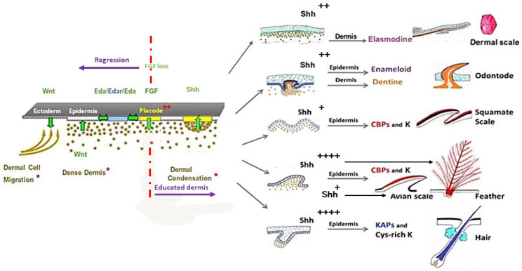
All living jawed vertebrates possess teeth or did so ancestrally. Integumental surface also includes the cornea. Conversely, no other anatomical feature differentiates the clades so readily as skin appendages do, multicellular glands in amphibians, hair follicle/gland complexes in mammals, feathers in birds, and the different types of scales. Tooth-like scales are characteristic of chondrichthyans, while mineralized dermal scales are characteristic of bony fishes. Corneous epidermal scales might have appeared twice, in squamates, and on feet in avian lineages, but posteriorly to feathers. In contrast to the other skin appendages, the origin of multicellular glands of amphibians has never been addressed. In the seventies, pioneering dermal-epidermal recombination between chick, mouse and lizard embryos showed that: (1) the clade type of the appendage is determined by the epidermis; (2) their morphogenesis requires two groups of dermal messages, first for primordia formation, second for appendage final architecture; (3) the early messages were conserved during amniotes evolution. Molecular biology studies that have identified the involved pathways, extending those data to teeth and dermal scales, suggest that the different vertebrate skin appendages evolved in parallel from a shared placode/dermal cells unit, present in a common toothed ancestor, c.a. 420 mya.

Keywords: cornea; development; evolution; feather; hair; placode; reticula; scale; tooth.


Conflict of interest statement

The author declares no conflict of interest.

Figures

Figure 1
Figure 1
In mammals, a balance between hair and cornea, as well as between hair and teeth. (A,A’,B,B’) In mutant mice, when Dkk2, a Wnt signaling inhibitor, is ablated, the cornea is transformed into a hairy skin. h: hair. (C,C’) A similar phenotype was observed in a wild deer with hair covering its cornea. (D) A transient tooth bud in a bowhead whale fetus: iee: inner enamel epithelium, oee: outer enamel epithelium. (E) Subgingival hair associated with a canine in a Labrador retriever dog. Credits: (A,A’,B,B’): After Mukhopadhyay et al. 2006, [35], reprinted with permission of the Company of Biologists. (C,C’): By courtesy of Lindsay Thomas, Chief Communications of the National Deer Association (USA). (D) After Thewissen et al. 2017 [36], reprinted with permission of John Wiley and sons. (E) By courtesy of Jan Bellows, medical director at All Pets Dental in Weston, Florida (USA).
Figure 2
Figure 2
Terrestrial life has been promoted by the development of plantar domains, regulated by the same Shh/BMP balance, both in bird lineage and mammals. (A) Basal ornithischian dinosaur Kulindadromeus shows plantar reticula. (B) In chick embryos, the treatment by retinoic acid at E11 enhances the amount of Shh in skin and leads at E17 to the formation of feathers instead of numerous reticula. (C1C3) In mice, the overexpression of Noggin under K14 promoter inhibits BMP4, leading to the formation of hair follicles in the foot pads. Schema: It should be noted that the amount of Shh expression in overlapping scale of birds is intermediate between feather (high) and reticula (low). ESC: epidermal scale, HFG: hair follicle gland complex, PF: protofeather, F: feather. Credits: (A) by courtesy of P. Godefroit, (B) image Dhouailly, (C1–C3) After Plikus et al., 2004, [57], reprinted with permission from Elsevier. Schema: Dhouailly.
Figure 3
Figure 3
In contrast to the evolution of feather, hair and squamate scale morphogenesis appear to have not particularly varied. Squamates originate in Late Triassic, and we can presume that they were covered by epidermal scales. Fossils of pterosaurs and early dinosaurs indicate that they were fluffy animals, so we can presume that single barbs were present in their common ancestor, the ornithodires. The evolution of feather architecture appears to not follow the same path, and the rachis formation can precede or not the formation of the barbules. Moreover, several types of feathers do not exist as the ribbon feathers of Epidexipteryx anymore. In modern mammals, hair has a simple architecture which was already present in Early Cretaceous mammalia. Credits: Kulindadromeus by courtesy of P. Godefroit, Anurognathid by courtesy of M. Benton, Spinolestes, by courtesy of T. Martin. Schema of a ribbon like feather of Epidexipteryx, modified after Xu and Guo [81]. Schema Dhouailly.
Figure 4
Figure 4
Two major steps in feather and hair morphogenesis, but only one in squamate scale. Xenoplastic dermal epidermal recombinants cultured on chick chorioallantoïc membrane. Note the darkly staining of mouse nuclei. (A1) E7 chick epidermis recombined with E14.5 mouse dermis leads after 4 days of culture to the formation of feather placodes associated to mouse dermal condensation. (A2) After 8 days of culture, abnormal short feathers are observed with recognizable alignment of cells (A3), characteristic of barb ridges. (B1) Recombinant of E12 mouse epidermis and E7 chick dermis leads after 3 days to formation of hair placodes associated to chick dermal condensation then (B2) to hair buds still associated to a chick dermal condensation. (B3) However, after 8 days of culture the dermal papilla has been dispersed, and despite an elongation of the hair bud, the hair morphogenesis was interrupted. (C) Recombinant of E 17 lizard epidermis and E14.5 mouse dermis leads, after 8 days, to the formation of scales with their corneous layer. Credits: After Dhouailly D, 1973, [102] and Dhouailly D, 1975, [103].
Figure 5
Figure 5
Different placodal shapes revealed by the expression of markers in different vertebrates. (A) Oval dental placodes of incisors of E12 mouse, as revealed by Shh expression. (B1,B2) Round femoral feather placodes of E7.5 chick as revealed by EDA in the inter-placodal and EDAR in the placodes. (C) Round dorsal hair placodes of E14.5 mouse, as revealed by Shh expression. (D1,D2) Crescent placodes of a 9–10 mm fry zebrafish, as revealed by the expression of EDA in the inter-placodal (ip) and of EDAR in the placodes (p). (E) Round placodes of plantar reticula of a E14 chick are revealed by the expression of EN1. The arrow indicates the central foot pad. (F) Round dorsal and rectangular ventral placodes of a E26 snake are revealed by Shh expression. Credits: (A) by Courtesy of Dr. I. Thesleff; (B1,B2) After Houghton et al., 2005, [134], reprinted with permission of the company of biologists; (C) After Huang et al., 2012 [135]; (D1,D2) by courtesy of Dr. M. Harris, Harris et al., 2008 [64]. (E) After. Prin and Dhouailly, 2004 [55]. (F) from the work of Di-Po and Milikovitch, 2016 [120].
Figure 6
Figure 6
When EDA or EDAR are missing, the results are identical in Zebrafish and in Humans. Cutaneous appendages morphogenesis is deficient in absence of EDA/EDAR signaling. Rare dermal scales in Zebrafish (compare figures A1, B1), with a total absence of teeth (compare figures A2, B2). Rare hair (C1) and teeth (C2) in Human. Credit: by courtesy of M. Harris, 2008 [64].
Figure 7
Figure 7
The first group of developmental pathways, leading to cutaneous appendage buds, is common to all vertebrates. The absence of Wingless-integrated (Wnt) prevents dermis formation and placode initiation in all types of appendages in all species. The EDA/EDAR pathway, activated downstream of Wing signaling, triggers several pathways, among others FGF and Shh, which are required both for the formation of the dermal condensation and the growth of the placode (**), respectively. FGF plays a pivotal role (see text). A second group of pathways governing the specific architecture of each kind of cutaneous appendage varies, not in type, but in location and timing, mostly Wnts, Shh, BMPs, Noggin, Gremlin. The regulation of Shh is of critical importance for the growth of the different appendages, from low (+), to medium (++), to very high (++++). Note that the simple architecture of squamate epidermal scales does not involve an elaborate second group of messages. * Three steps of dermis formation. Note that dermal condensation does not exist in squamates. CBP: corneous beta proteins; K: keratins, KAPs: keratin associated proteins, cys-rich K: cysteine rich keratins. Schema: Dhouailly.

Similar articles

Cited by

References

    1. Chammas S.M., Carneiro S.M., Ferro R.S., Antoniazzi M.M., Jared C. Development of integument and cutaneous glands in larval, juvenile and adult toads (Rhinella granulosa): A morphological and morphometric study. Acta Zool. 2015;96:460–477. doi: 10.1111/azo.12091. - DOI
    1. Çömden E.A., Yenmiş M., Çakır B. The Complex Bridge between Aquatic and Terrestrial Life: Skin Changes during Development of Amphibians. J. Dev. Biol. 2023;11:6. doi: 10.3390/jdb11010006. - DOI - PMC - PubMed
    1. Hirasawa T., Nagashima H., Kuratani S. The endoskeletal origin of the turtle carapace. Nat. Commun. 2013;4:2107. doi: 10.1038/ncomms3107. - DOI - PMC - PubMed
    1. Chen I.H., Yang W., Meyers M.A. Alligator osteoderms: Mechanical behavior and hierarchical structure. Mater. Sci. Eng. C. 2014;35:441–448. doi: 10.1016/j.msec.2013.11.024. - DOI - PubMed
    1. Lucas A.M., Stettenheim P.R. Avian Anatomy Integument. US Department of Agriculture; Washington, DC, USA: 1972.

LinkOut - more resources