. 2023 Jul 27;97(7):e0068623.
doi: 10.1128/jvi.00686-23. Epub 2023 Jun 27.

Foot-and-Mouth Disease Virus Induces Porcine Gasdermin E-Mediated Pyroptosis through the Protease Activity of 3Cpro

Affiliations

Foot-and-Mouth Disease Virus Induces Porcine Gasdermin E-Mediated Pyroptosis through the Protease Activity of 3Cpro

Xujiao Ren et al. J Virol. .

Abstract

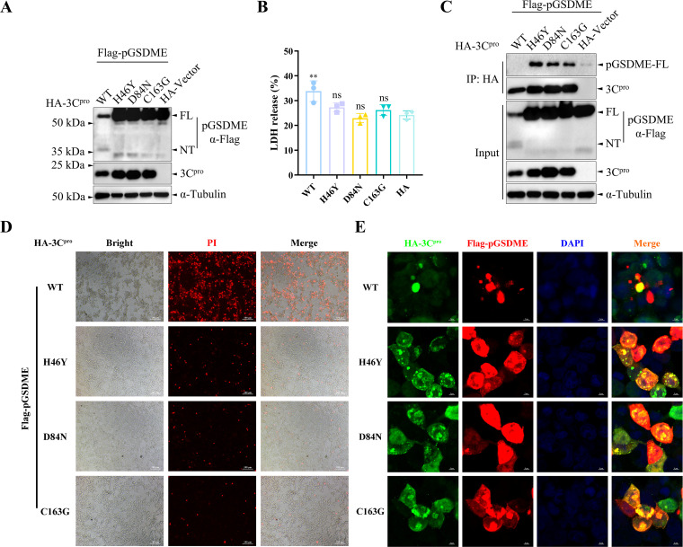
Foot-and-mouth disease (FMD) is an acute, highly contagious disease of cloven-hoofed animals caused by FMD virus (FMDV). Currently, the molecular pathogenesis of FMDV infection remains poorly understood. Here, we demonstrated that FMDV infection induced gasdermin E (GSDME)-mediated pyroptosis independent of caspase-3 activity. Further studies showed that FMDV 3Cpro cleaved porcine GSDME (pGSDME) at the Q271-G272 junction adjacent to the cleavage site (D268-A269) of porcine caspase-3 (pCASP3). The inhibition of enzyme activity of 3Cpro failed to cleave pGSDME and induce pyroptosis. Furthermore, overexpression of pCASP3 or 3Cpro-mediated cleavage fragment pGSDME-NT was sufficient to induce pyroptosis. Moreover, the knockdown of GSDME attenuated the pyroptosis caused by FMDV infection. Our study reveals a novel mechanism of pyroptosis induced by FMDV infection and might provide new insights into the pathogenesis of FMDV and the design of antiviral drugs. IMPORTANCE Although FMDV is an important virulent infectious disease virus, few reports have addressed its relationship with pyroptosis or pyroptosis factors, and most studies focus on the immune escape mechanism of FMDV. GSDME (DFNA5) was initially identified as being associated with deafness disorders. Accumulating evidence indicates that GSDME is a key executioner for pyroptosis. Here, we first demonstrate that pGSDME is a novel cleavage substrate of FMDV 3Cpro and can induce pyroptosis. Thus, this study reveals a previously unrecognized novel mechanism of pyroptosis induced by FMDV infection and might provide new insights into the design of anti-FMDV therapies and the mechanisms of pyroptosis induced by other picornavirus infections.

Keywords: 3Cpro; cleavage; foot-and-mouth disease virus; porcine gasdermin E; protease activity; pyroptosis.


Conflict of interest statement

The authors declare no conflict of interest.

Figures

FIG 1
FIG 1
FMDV infection induces pyroptosis in SK6 cells. SK6 cells were infected with FMDV O/HN/CHA/93 strain at an MOI of 1 at the indicated times. (A) Morphological examination of SK6 cells with FMDV or mock infection. SVV infection was used as a positive control. The cell morphology was observed using an Olympus IX71 microscope (white arrows, pyroptotic cells). (B) FMDV inhibited SK6 cell viability. The cell viability of SK6 was determined by using a Cell Counting Kit-8. (C) SK6, PK-15, BHK-21, and HEK-293T cells were infected with FMDV O/HN/CHA/93 strain at an MOI of 1 at the indicated times. The release of LDH from FMDV-infected cells was measured with an LDH cytotoxicity assay kit. (D) PI staining of SK6 cells with FMDV or mock infection. (E and F) The relative expression levels of IL-1β and IL-18 were analyzed by quantitative RT-PCR (qRT-PCR) in SK6 cells, and the housekeeping gene GAPDH was used as the control. (G) FMDV infection induces GSDME-mediated pyroptosis in SK6 cells. Proteolytic cleavage of GSDMD or GSDME in SK6 cells with FMDV infection was determined by Western blotting. The abundance of caspase-3 (CASP3), IL-1β, MLKL, phospho-MLKL, FMDV VP1 protein, and α-tubulin as an internal control was also determined by Western blotting. Two lanes represent two biological replicates. (H and I) The release of IL-1β and IL-18 from FMDV-infected cells was measured with porcine IL-1β and IL-18 using an enzyme-linked immunosorbent assay (ELISA) kit. (J and K) SK6 cells were transfected with plasmids encoding pGSDME for 24 h and then infected with FMDV O/HN/CHA/93 strain at an MOI of 1 at the indicated times. (L and M) SK6 cells were infected with FMDV at various MOIs as indicated for 24 h. Proteolytic cleavage of GSDME in SK6 cells with FMDV infection was determined by Western blotting. The release of LDH from FMDV-infected cells was measured with LDH Cytotoxicity assay kit. The data in panels B, C, E, F, H, I, K, and M are presented as means ± the SD. Experiments were performed independently with at least two biological replicates. Student t test or one/two-way ANOVA was used for analysis. *, P < 0.05; **, P < 0.01; ***, P < 0.001; ****, P < 0.0001.
FIG 2
FIG 2
FMDV infection induces pyroptosis in SK6 cells independent of caspase-3 activity. SK6 cells were infected with FMDV O/HN/CHA/93 strain at an MOI of 1 for 12 h in the presence or absence of Ac-DEVD-CHO (20 μM). (A) Morphological examination of SK6 cells with FMDV or mock infection. The cell morphology was observed using an Olympus IX71 microscope (white arrows, pyroptotic cells). (B) The release of LDH from FMDV-infected cells was measured with an LDH cytotoxicity assay kit. (C) PI staining of SK6 cells with FMDV or mock infection. (D) Proteolytic cleavage of GSDME in SK6 cells with FMDV infection was determined by Western blotting. The abundance of caspase-3 (CASP3), IL-1β, FMDV VP1 protein, and α-tubulin as an internal control was also determined by Western blotting. (E and F) The release of IL-1β and IL-18 from FMDV-infected cells was measured with porcine IL-1β and IL-18 using an ELISA kit. The data in panels B, E, and F are presented as means ± the SD. Experiments were performed independently with at least three biological replicates. Two-way ANOVA was used for analysis. ns, not significant.
FIG 3
FIG 3
FMDV 3Cpro induces pyroptosis in SK6 cells. (A) Proteolytic cleavage of pGSDME in SK6 cells by viral proteins. SK6 cells were cotransfected with plasmids encoding pGSDME and viral proteins for 24 h. Proteolytic cleavage of pGSDME in SK6 cells by viral proteins was determined by Western blotting. The red asterisk indicates the pGSDME N-terminal cleavage product. (B) SK6 cells were transfected with recombinant plasmids encoding 3Cpro. The cell morphology was observed using an Olympus IX71 microscope (white arrows, pyroptotic cells). (C) The release of LDH from 3Cpro-transfected cells was measured using an LDH cytotoxicity assay kit. (D) Proteolytic cleavage of pGSDME in SK6 cells by 3Cpro. SK6 cells were cotransfected with plasmids encoding 3Cpro and pGSDME for 24 h. Proteolytic cleavage of pGSDME in SK6 cells by 3Cpro was determined by Western blotting. Two lanes represent two biological replicates. The data in panel C are presented as means ± the SD. Experiments were performed independently with at least three biological replicates. A Student t test was used for analysis. *, P < 0.05.
FIG 4
FIG 4
Catalytic residues in the active sites of 3Cpro are essential for the pGSDME cleavage induced by 3Cpro. (A) The effects of catalytic residues of the 3Cpro active sites on 3Cpro-induced pGSDME cleavage. HEK-293T cells were cotransfected with plasmids encoding pGSDME and 3Cpro or its mutants for 12 h. Proteolytic cleavage of pGSDME in HEK-293T cells by 3Cpro or its mutants was determined by Western blotting. (B) The release of LDH from pGSDMD and 3Cpro-transfected cells was measured with an LDH cytotoxicity assay kit. (C) FMDV 3Cpro interacts with and cleaves pGSDME. HEK-293T cells were cotransfected with plasmids encoding pGSDME and 3Cpro or its mutants for 12 h. The interaction between 3Cpro and pGSDME was analyzed by using coimmunoprecipitation. (D) PI staining of the transfected cells. (E) The interaction between 3Cpro and pGSDME was analyzed by using confocal immunofluorescence. The data in panel B are presented as means ± the SD. Experiments were performed independently with at least three biological replicates. One-way ANOVA was used for analysis. **, P < 0.01.
FIG 5
FIG 5
Rupintrivir inhibits FMDV-induced pyroptosis in SK6 cells. SK6 cells were infected with FMDV O/HN/CHA/93 strain at an MOI of 1 for 12 h in the presence or absence of rupintrivir (8 μM). (A) Chemical formula of rupintrivir (AG7088). (B) Binding mode of rupintrivir to FMDV 3Cpro. Molecular docking studies were performed by Autodock Vina 1.2.2 (http://autodock.scripps.edu/) and visualized using the PyMOL 2.5 software (DeLano Scientific LLC), with PDB ID 5hm2 (FMDV 3Cpro) and 4ght (EV71 3Cpro-AG7088 complex) used as the templates. The yellow structure represents rupintrivir, and the pink residues represent His46 (H46) and Asp84 (D84). (C) Rupintrivir inhibits the activity of 3Cpro in a dose-dependent manner. HEK-293T cells were cotransfected with plasmids encoding 3Cpro and pGSDME for 12 h in the presence or absence of rupintrivir. Proteolytic cleavage of pGSDME in HEK-293T cells by 3Cpro was determined by Western blotting. (D) PI staining of SK6 cells with FMDV or mock infection. (E) The release of LDH from FMDV-infected cells was measured with an LDH cytotoxicity assay kit. (F) Proteolytic cleavage of GSDME in SK6 cells with FMDV infection was determined by Western blotting. The abundance of caspase-3 (CASP3), IL-1β, FMDV VP1 protein, and α-tubulin as an internal control was also determined by Western blotting. (G and H) The release of IL-1β and IL-18 from FMDV-infected cells was measured with porcine IL-1β and IL-18 using an ELISA kit. The data in panels E, G, and H are presented as means ± the SD. Experiments were performed independently with at least three biological replicates. Two-way ANOVA was used for analysis. ****, P < 0.0001.
FIG 6
FIG 6
FMDV 3Cpro cleaves pGSDME adjacent to the cleavage site of pCASP3. (A) Sequence alignment of GSDME in pigs, humans, and mice. (B) Logo analysis of the cleavage sites predicted from the polyprotein cleavage of pCaspase-3 (pCASP3) or 3Cpro. (C) Cleavage sites of pGSDME induced by pCASP3. HEK-293T cells were cotransfected with plasmids encoding pCASP3 and pGSDME or its mutants for 24 h in the presence or absence of TNF-α (50 ng/mL). (D) Cleavage sites of pGSDME induced by 3Cpro. HEK-293T cells were cotransfected with plasmids encoding 3Cpro and pGSDME or its mutants for 12 h. Proteolytic cleavage of pGSDME or its mutants in HEK-293T cells by pCASP3 or 3Cpro was determined by Western blotting.
FIG 7
FIG 7
pCASP3 or 3Cpro -mediated cleavage fragment pGSDME-NT localizes to the cell membrane and triggers pyroptosis. (A) Schematic diagram of pGSDME and its cleavage fragments induced by pCaspase-3 (pCASP3) or 3Cpro. HEK-293T cells were transfected with recombinant plasmids encoding pGSDME or its cleavage fragments for 48 h. (B) The expression of pGSDME and its cleavage fragments was determined by Western blotting. (C) The localization of pGSDME or its cleavage fragments was analyzed by confocal immunofluorescence. (D) The release of LDH from the transfected cells was measured with an LDH cytotoxicity assay kit. (E) PI staining of the transfected cells. (F) The cell morphology was observed using an Olympus IX71 microscope (white arrows, pyroptotic cells). The data in panel D are presented as means ± the SD. Experiments were performed independently with at least three biological replicates. One-way ANOVA was used for analysis. ****, P < 0.0001.
FIG 8
FIG 8
pGSDME knockdown attenuates FMDV-induced pyroptosis in SK6 cells. SK6 cells were transduced with lentivirus targeting pGSDME (sh-pGSDME) or negative control (sh-NC) for 24 h, and then the cells were infected with FMDV O/HN/CHA/93 strain at an MOI of 1 for 12 h. (A) The knockdown efficiency of pGSDME was determined by Western blotting. (B) Morphological examination of SK6 cells with FMDV or mock infection. The cell morphology was observed using an Olympus IX71 microscope (white arrows, pyroptotic cells). (C) The release of LDH from FMDV-infected cells was measured with an LDH cytotoxicity assay kit. (D) PI staining of SK6 cells with FMDV or mock infection. (E) Proteolytic cleavage of GSDME in SK6 cells with FMDV infection was determined by Western blotting. The abundance of caspase-3, IL-1β, FMDV VP1 protein, and α-tubulin as an internal control was also determined by Western blotting. (F and G) The release of IL-1β and IL-18 from FMDV-infected cells was measured with porcine IL-1β and IL-18 using an ELISA kit. The data in panels C, F, and G are presented as means ± the SD. Experiments were performed independently with at least three biological replicates. Two-way ANOVA was used for analysis. *, P < 0.05; ***, P < 0.001.
FIG 9
FIG 9
Schematic diagram showing the proposed mechanism of FMDV-induced GSDME-mediated pyroptosis. FMDV infection induces GSDME-mediated pyroptosis independent of caspase-3 activity. In addition, FMDV 3Cpro induces pyroptosis by cleaving pGSDME adjacent to the cleavage site of pCaspase-3, and the protease activity of 3Cpro is required for pGSDME cleavage.

References

    1. David W, Brown G. 2001. Foot and mouth disease in human beings. Lancet 357:1463. doi:10.1016/S0140-6736(00)04670-5. - DOI - PubMed
    1. Thomson GR, Vosloo W, Bastos AD. 2003. Foot and mouth disease in wildlife. Virus Res 91:145–161. doi:10.1016/s0168-1702(02)00263-0. - DOI - PubMed
    1. Sangare O, Bastos AD, Marquardt O, Venter EH, Vosloo W, Thomson GR. 2001. Molecular epidemiology of serotype O foot-and-mouth disease virus with emphasis on West and South Africa. Virus Genes 22:345–351. doi:10.1023/a:1011178626292. - DOI - PubMed
    1. Grubman MJ, Baxt B. 2004. Foot-and-mouth disease. Clin Microbiol Rev 17:465–493. doi:10.1128/CMR.17.2.465-493.2004. - DOI - PMC - PubMed
    1. Curry S, Roqué-Rosell N, Zunszain PA, Leatherbarrow RJ. 2007. Foot-and-mouth disease virus 3C protease: recent structural and functional insights into an antiviral target. Int J Biochem Cell Biol 39:1–6. doi:10.1016/j.biocel.2006.07.006. - DOI - PMC - PubMed

Publication types

LinkOut - more resources