Review
. 2023 Jun 14;21(6):359.
doi: 10.3390/md21060359.

Cyanobacteria: A Promising Source of Antifungal Metabolites

Affiliations
Review

Cyanobacteria: A Promising Source of Antifungal Metabolites

Samuel Cavalcante do Amaral et al. Mar Drugs. .

Abstract

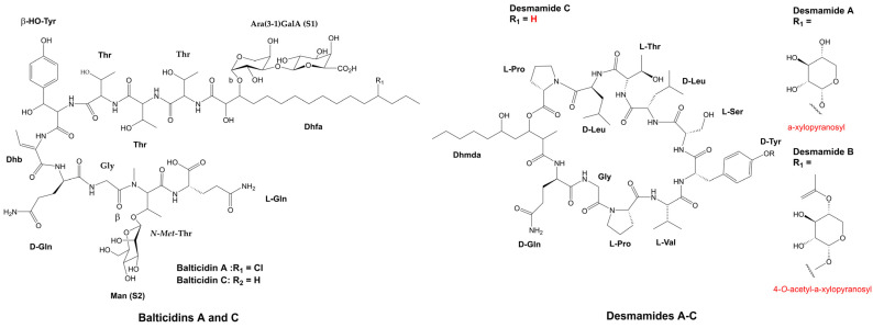
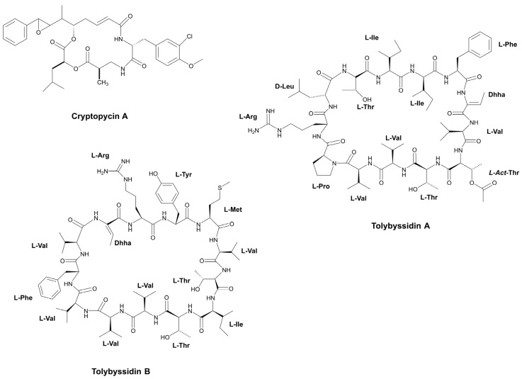
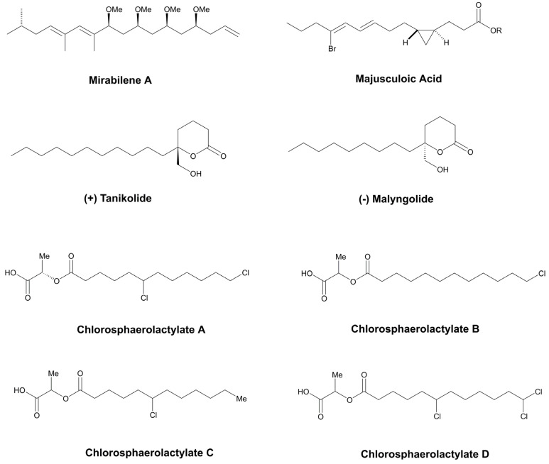
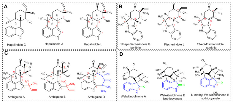
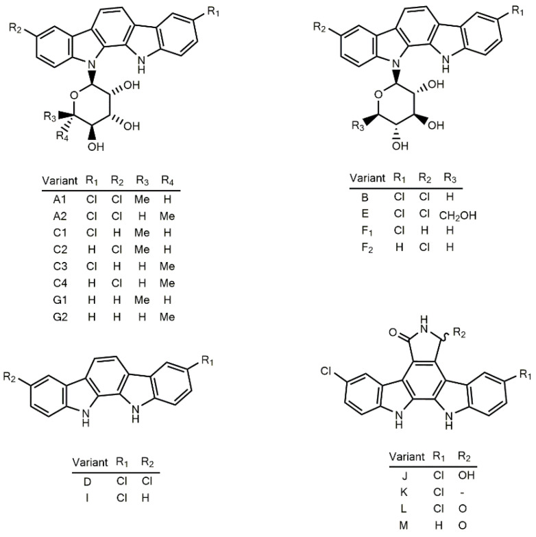
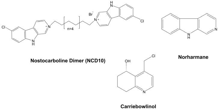
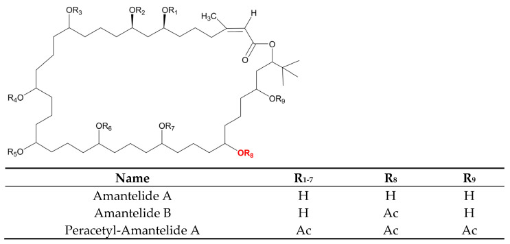
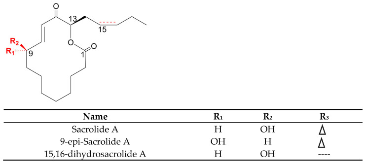
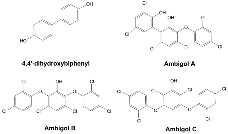
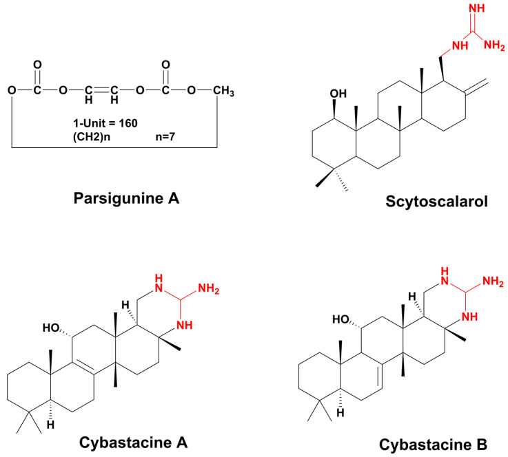
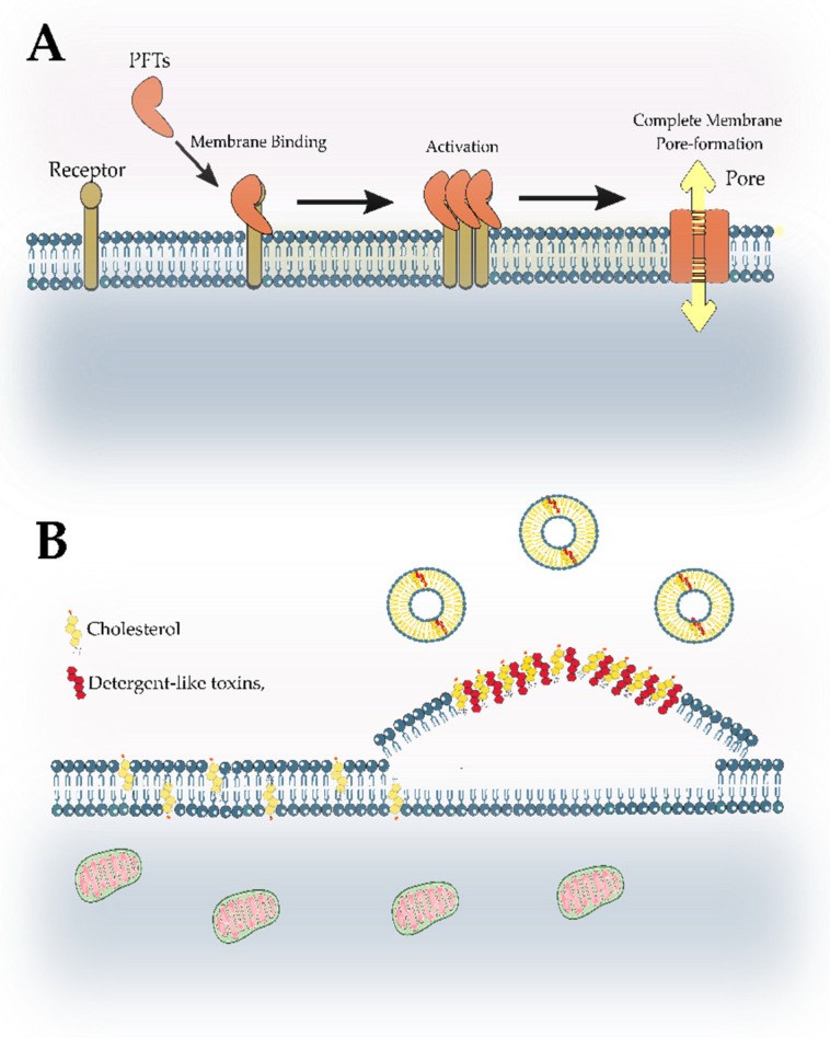
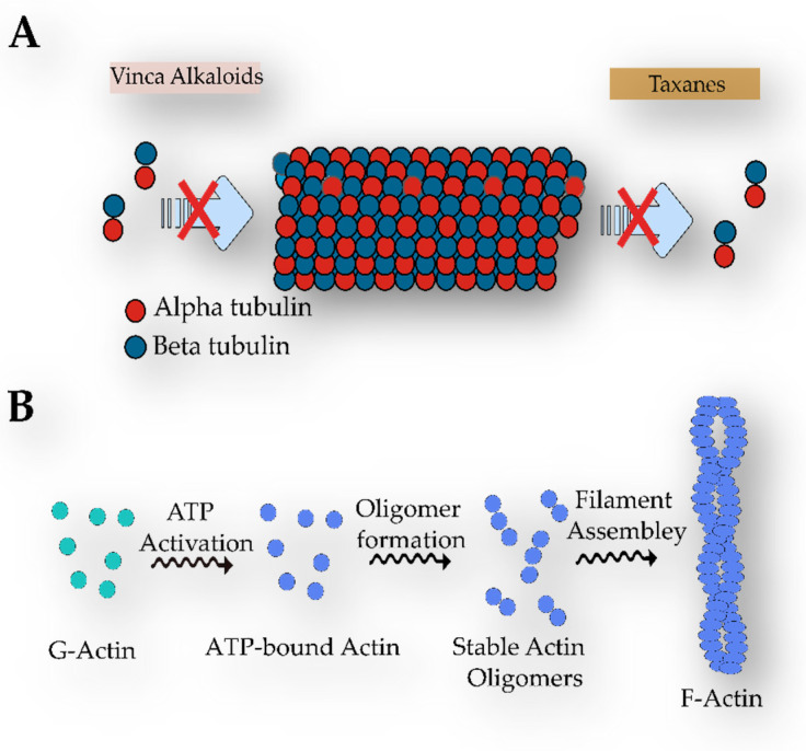
Cyanobacteria are a rich source of secondary metabolites, and they have received a great deal of attention due to their applicability in different industrial sectors. Some of these substances are known for their notorious ability to inhibit fungal growth. Such metabolites are very chemically and biologically diverse. They can belong to different chemical classes, including peptides, fatty acids, alkaloids, polyketides, and macrolides. Moreover, they can also target different cell components. Filamentous cyanobacteria have been the main source of these compounds. This review aims to identify the key features of these antifungal agents, as well as the sources from which they are obtained, their major targets, and the environmental factors involved when they are being produced. For the preparation of this work, a total of 642 documents dating from 1980 to 2022 were consulted, including patents, original research, review articles, and theses.

Keywords: action mechanism; alkaloids; antifungal agents; cyanobacteria; peptides; secondary metabolites.


Conflict of interest statement

The authors declare no conflict of interest.

Figures

Figure 1
Figure 1
The number of metabolites with antifungal properties identified and shared among strains of cyanobacteria from different environments, including environmental samples.
Figure 2
Figure 2
Percentage of compound in each genus.
Figure 3
Figure 3
Chemical structure of some antifungal cyanopeptides with β-amino acids.
Figure 4
Figure 4
Similarity (red) and differences between the members of the Puwainaphycin family and the cyclopeptide, Calophycin.
Figure 5
Figure 5
Fungicide isolated from cyanobacteria. (A) Nostofungicidine. (B) Anabaenolysins A and B. (C) Cyclodextrin variants at different acetylation levels. (D) Laxaphycins A and E. (E) Hormothamnin A. (F) Laxaphycins B and D.
Figure 6
Figure 6
Structure of the peptides, Microcolins A and B.
Figure 7
Figure 7
Biosynthesis of oxazole and thiazole rings from the amino acids, Serine and Cysteine, respectively.
Figure 8
Figure 8
Cyanopeptides with fungicidal properties bearing thiazole and oxazole rings.
Figure 9
Figure 9
The structure of Hassallidins A–E. Abbreviations: Dhfa (dihydroxy fatty acid), dihydroxy tetradecanoic acid in hassallidin A and B, and dihydroxy hexadecanoic acid in hassallidin C, D, and E.
Figure 10
Figure 10
Chemical structure of Balticidin A and C, and the recently discovered Desmamides A–C.
Figure 11
Figure 11
Example of fungicidal cyanopeptides released or excreted into the supernatant. Abbreviations: L-Arg (L-arginine), L-Tyr (L-tyrosine), L-Met (L-methionine), L-Val (L-valine), L-Thr (L-threonine), L-Ile (L-isoleucine), L-Phe (L-phenylalanine), Dhha (dehydrohomoalanine), L-Pro (L-proline), D-Leu (D-leucine), O-Ac-Thr (O-Acetyl-L-Threonine).
Figure 12
Figure 12
Fungal cell wall components. The fungal cell wall is composed of a membrane enriched with ergosterol (in yellow) and some proteins. It is also possible to observe the presence of a protective layer of chitin, as well as glucans, and mannoproteins on the surface.
Figure 13
Figure 13
Hydrolysis of chitosan by the enzyme chitosanase (Cho), which is produced and released by the strain, Anabaena fertilissima RPAN1.
Figure 14
Figure 14
Fatty acids and their derivatives which possess fungicidal activity, isolated from cyanobacteria.
Figure 15
Figure 15
Hapalindole-type alkaloids. (A). Some examples of tricycle hapalindoles (Hapalindole C) and tetracyclo hapalindoles (J and C). In the tetracyclic hapalindoles, the key connection occurs between C-4 and C-16. (B). Chemical structure of some Fischerindoles, in which the key connection is between C-2 and C-16 (C). Chemical structure of Ambiguines A, B, and C. In red, the isoprene unit is attached to C-2, and in blue, the isoprenyl group is fused to the isonitrile-bearing carbon. (D) Some examples of welwitindoliones. In green, the oxidized carbon is in the second position, and in blue, the bicyclo is shown [4.3.1].
Figure 16
Figure 16
Chemical structure of members of the Tjipanazoles family.
Figure 17
Figure 17
Chemical structure of alkaloids, with fungicidal properties, obtained from cyanobacteria.
Figure 18
Figure 18
Chemical structure of the Polyketides, Kalkipyrones A–B, and Yoshinone. In red, the difference between Kalkipyrone A and Yoshinone is shown.
Figure 19
Figure 19
Carbamidocyclophanes’ (A–F) chemical structures.
Figure 20
Figure 20
Chemical structure of Scytophycins.
Figure 21
Figure 21
Chemical structure of the macrolides Swinholide A and Ankaraholides A–B.
Figure 22
Figure 22
Chemical structure of Amantelides A–B and the Peracetyl-Amantelide A.
Figure 23
Figure 23
Structure of Sacrolide A and its congeners 9-epo-Sacrolide A and 15,16-dihydrosacrolide A.
Figure 24
Figure 24
Phenolic compounds with antifungal activity of cyanobacterial origin.
Figure 25
Figure 25
Structure of the polymer Parsiguine A, terpenoid Scytoscalarol, and Cybastacines A–B. In red, the guanidine group is shown.
Figure 26
Figure 26
Molecular mechanisms of membrane-targeting compounds. (A) Pathways of pore-forming toxins (PFTs). Soluble PFTs move to plasmatic membrane and bind to receptors molecules. Then, they oligomerize on the surface of the membrane and produce transmembrane pores. (B) General mechanism of detergent-like toxins. These molecules normally bind to the external monolayer of the plasmatic membrane containing cholesterol and promote the vesicles formation and lateral phase separation.
Figure 27
Figure 27
Structure of a Microtubule and the Actin Filaments. (A) Mechanism of action of vinca alkaloids and taxanes. Although taxanes promote microtubule stabilization by avoiding the release of dimers of tubulin, vinca alkaloids block microtubule polymerization. (B) F-actin formation.
Figure 28
Figure 28
Light parameters normally investigated in studies concerning cyanobacterial growth and the production of secondary metabolites. (A) Light intensity, (B) Photoperiod, (C) Wavelength.

Similar articles

Cited by

References

    1. Kendrick B. Fungi: Ecological Importance and Impact on Humans. eLS. 2011:1–5. doi: 10.1002/9780470015902.a0000369.pub2. - DOI
    1. Friedman D.Z.P., Schwartz I.S. Emerging Fungal Infections: New Patients, New Patterns, and New Pathogens. J. Fungi. 2019;5:67. doi: 10.3390/jof5030067. - DOI - PMC - PubMed
    1. Lockhart S.R., Guarner J. Emerging and Reemerging Fungal Infections. Semin. Diagn. Pathol. 2019;36:177–181. doi: 10.1053/j.semdp.2019.04.010. - DOI - PMC - PubMed
    1. Jenks J.D., Cornely O.A., Chen S.C.A., Thompson G.R., Hoenigl M. Breakthrough Invasive Fungal Infections: Who Is at Risk? Mycoses. 2020;63:1021–1032. doi: 10.1111/myc.13148. - DOI - PubMed
    1. Garcia-Solache M.A., Casadevall A. Global Warming Will Bring New Fungal Diseases for Mammals. mBio. 2010;1:e00061-10. doi: 10.1128/mBio.00061-10. - DOI - PMC - PubMed

MeSH terms

LinkOut - more resources