. 2023 Jun 16;21(6):360.
doi: 10.3390/md21060360.

Antioxidant Peptides from Monkfish Swim Bladders: Ameliorating NAFLD In Vitro by Suppressing Lipid Accumulation and Oxidative Stress via Regulating AMPK/Nrf2 Pathway

Affiliations

Antioxidant Peptides from Monkfish Swim Bladders: Ameliorating NAFLD In Vitro by Suppressing Lipid Accumulation and Oxidative Stress via Regulating AMPK/Nrf2 Pathway

Ming-Feng Wu et al. Mar Drugs. .

Abstract

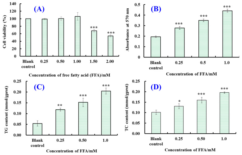
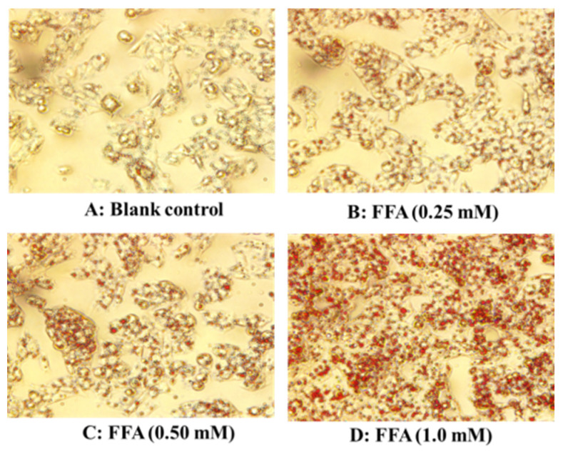
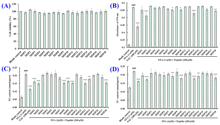
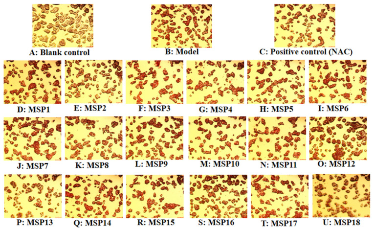
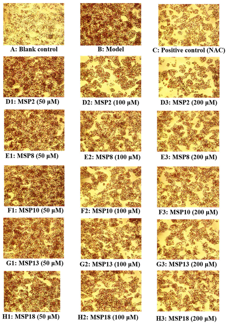
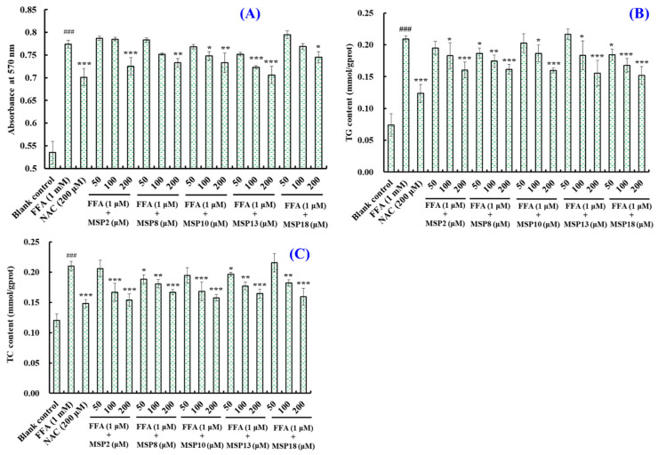
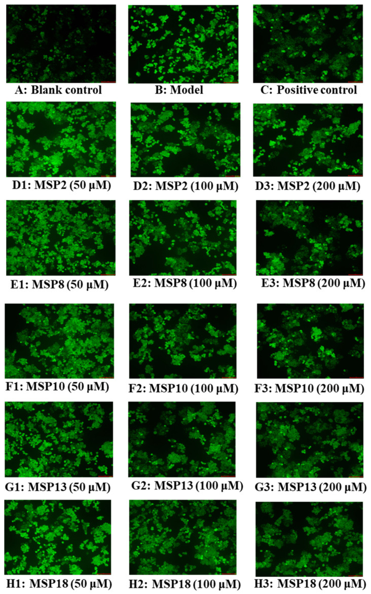
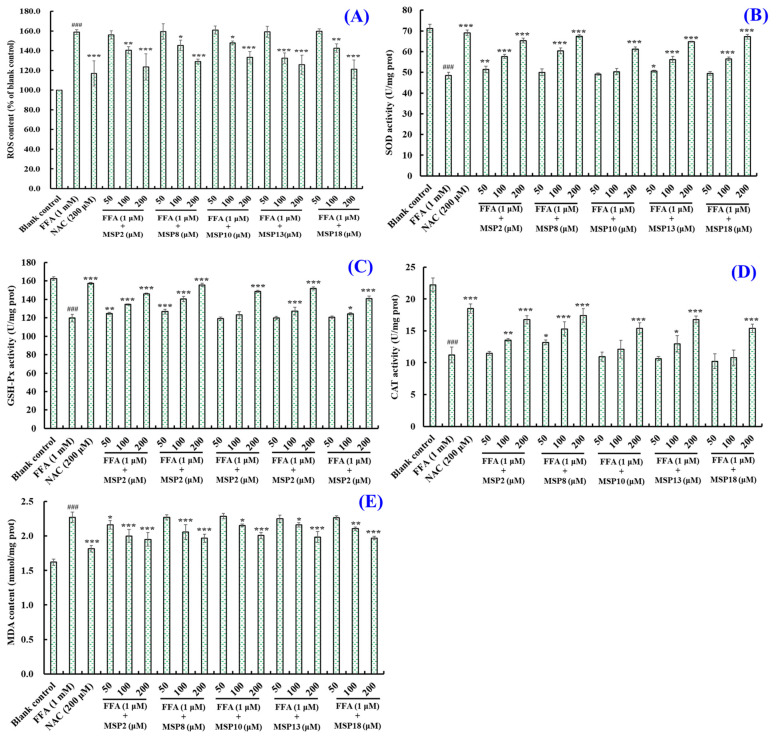
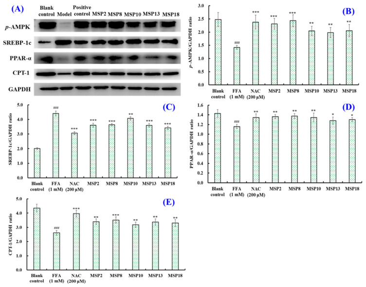
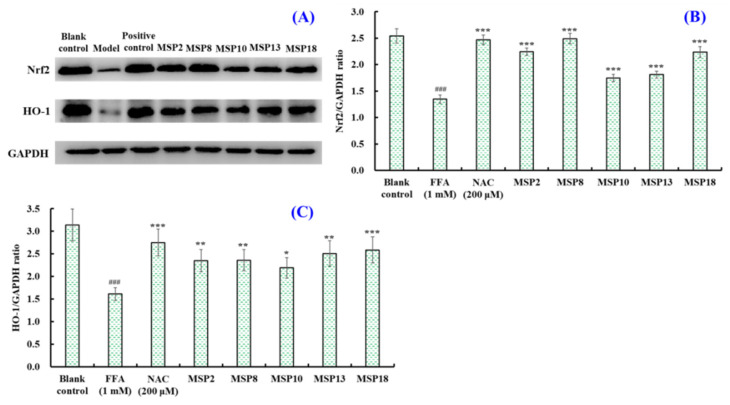
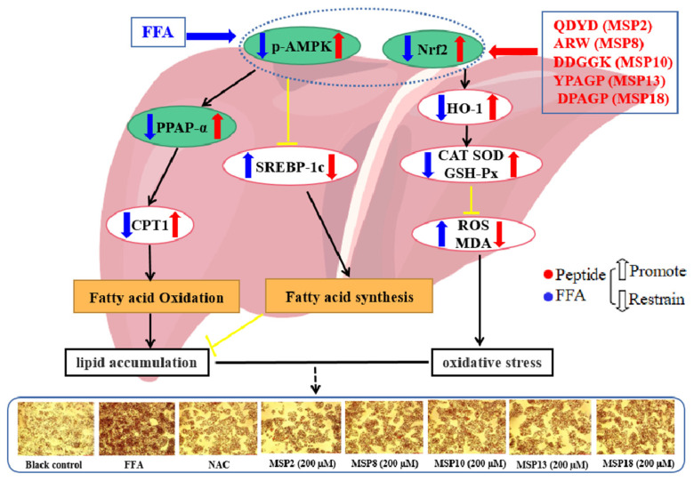
In this study, we investigate the ameliorating functions of QDYD (MSP2), ARW (MSP8), DDGGK (MSP10), YPAGP (MSP13) and DPAGP (MSP18) from monkfish swim bladders on an FFA-induced NAFLD model of HepG2 cells. The lipid-lowering mechanisms revealed that these five oligopeptides can up-regulate the expression of phospho-AMP-activated protein kinase (p-AMPK) proteins to inhibit the expression of the sterol regulatory element binding protein-1c (SREBP-1c) proteins on increasing lipid synthesis and up-regulating the expression of the PPAP-α and CPT-1 proteins on promoting the β-oxidation of fatty acids. Moreover, QDYD (MSP2), ARW (MSP8), DDGGK (MSP10), YPAGP (MSP13) and DPAGP (MSP18) can significantly inhibit reactive oxygen species' (ROS) production, promote the activities of intracellular antioxidases (superoxide dismutase, SOD; glutathione peroxidase, GSH-PX; and catalase, CAT) and bring down the content of malondialdehyde (MDA) derived from lipid peroxidation. Further investigations revealed that the regulation of these five oligopeptides on oxidative stress was achieved through activating the nuclear factor erythroid 2-related factor 2 (Nrf2) pathway to raise the expression levels of the heme oxygenase 1 (HO-1) protein and downstream antioxidant proteases. Therefore, QDYD (MSP2), ARW (MSP8), DDGGK (MSP10), YPAGP (MSP13) and DPAGP (MSP18) could serve as candidate ingredients to develop functional products for treating NAFLD.

Keywords: AMPK/Nrf2 pathway; antioxidant peptide; monkfish (Lophius litulon); nonalcoholic fatty liver disease (NAFLD); swim bladders.


Conflict of interest statement

The authors declare no conflict of interest.

Figures

Figure 1
Figure 1
Influences of free fatty acids (FFA) on cell viability (A), intracellular lipid accumulation (B), triglyceride (TG) content (C) and total cholesterol (TC) content (D) in HepG2 cells. All data are presented as the mean ± SD of triplicate results. *** p < 0.001, ** p < 0.01 and * p < 0.05 vs. blank control group.
Figure 2
Figure 2
Effects of FFA (0–1.0 mM) on morphological characteristics of HepG2 cells (×20): (A) blank control; (B) FFA (0.25 mM); (C) FFA (0.50 mM); (D): FFA (1.0 mM).
Figure 3
Figure 3
Effects of eighteen antioxidant peptides (MSP1–MSP18) on the viability of HepG2 cells (A) and intracellular lipid accumulation (B), and the TG (C) and TC (D) contents in FFA-induced NAFLD model of HepG2 cells after 24 h of treatment. NAC at 200 µM was served as the positive control. All data are presented as the mean ± SD of triplicate results. ### p < 0.001 vs. blank control group; *** p < 0.001, ** p < 0.01 and * p < 0.05 vs. FFA-induced NAFLD model of HepG2 cells.
Figure 4
Figure 4
Effects of antioxidant peptides (MSP1–MSP18) on morphological characteristics of FFA-induced non-alcoholic fatty liver disease (NAFLD) model of HepG2 cells (20×). N-Acetyl-L-cysteine (NAC) was used as a positive control.
Figure 5
Figure 5
Effects of MSP2, MSP8, MSP10, MSP13 and MSP18 at 50, 100 and 200 µM on intracellular lipid accumulation in FFA-induced NAFLD model of HepG2 cells for 24 h (20×): (A) blank control; (B) FFA-induced cell model; (C) positive control (NAC); (D1D3) MSP2 with the concentrations of 50, 100 and 200 μM, respectively; (E1E3) MSP8 with the concentrations of 50, 100 and 200 μM, respectively; (F1F3) MSP10 with the concentrations of 50, 100 and 200 μM, respectively; (G1G3) MSP13 with the concentrations of 50, 100 and 200 μM, respectively; (H1H3) MSP18 with the concentrations of 50, 100 and 200 μM, respectively.
Figure 6
Figure 6
Effects of MSP2, MSP8, MSP10, MSP13 and MSP18 at 50, 100 and 200 µM on intracellular lipid accumulation (A), and the TG (B) and TC (C) contents in FFA-induced NAFLD model of HepG2 cells for 24 h. NAC at 200 µM was served as the positive control. All data are presented as the mean ± SD of triplicate results. ### p < 0.001 vs. blank control group; *** p < 0.001, ** p < 0.01 and * p < 0.05 vs. FFA-induced NAFLD model of HepG2 cells.
Figure 7
Figure 7
Determination of ROS levels in HepG2 cells by DCFH-DA staining (20×): (A) blank control; (B) FFA-induced cell model; (C) positive control (NAC); (D1D3) MSP2 with the concentrations of 50, 100 and 200 μM, respectively; (E1E3) MSP8 with the concentrations of 50, 100 and 200 μM, respectively; (F1F3) MSP10 with the concentrations of 50, 100 and 200 μM, respectively; (G1G3) MSP13 with the concentrations of 50, 100 and 200 μM, respectively; (H1H3) MSP18 with the concentrations of 50, 100 and 200 μM, respectively.
Figure 8
Figure 8
Effects of MSP2, MSP8, MSP10, MSP13 and MSP18 on ROS (A), SOD (B), GSH-PX (C), CAT (D) and MDA (E) levels of FFA-induced NAFLD model of HepG2 cells at 50, 100 and 200 µM. NAC at 200 µM was served as the positive control. All data are presented as the mean ± SD of triplicate results. ### p < 0.001 vs. blank control group; *** p < 0.001, ** p < 0.01 and * p < 0.05 vs. FFA-induced NAFLD model of HepG2 cells.
Figure 9
Figure 9
The effects of MSP2, MSP8, MSP10, MSP13 and MSP18 at a concentration of 200 μM on the expression of lipid metabolism-related proteins in FFA-induced NAFLD model in HepG2 cells. NAC at 200 µM was served as the positive control. All data are presented as the mean ± SD of triplicate results. (A) Expression levels of lipid metabolism related proteins; (B) p-AMPK/GAPDH ratio; (C) SREBP-1c/GAPDH ratio; (D) PPAR-α/GAPDH ratio; (E) CPT-1/GAPDH ratio. ### p < 0.001 vs. blank control group; *** p < 0.001, ** p < 0.01 and * p < 0.05 vs. FFA-induced NAFLD model of HepG2 cells.
Figure 10
Figure 10
The effects of MSP2, MSP8, MSP10, MSP13 and MSP18 at 200 μM on the expression of antioxidant-system-related proteins in FFA-induced NAFLD model in HepG2 cells. NAC at 200 µM was served as the positive control. All data are presented as the mean ± SD of triplicate results. (A) Expression levels of antioxidant-system-related proteins; (B) Nrf2/GAPDH ratio; (C) HO-1/GAPDH ratio. ### p < 0.05 vs. blank control group; *** p < 0.01, ** p < 0.01 and * p < 0.05 vs. FFA-induced NAFLD model of HepG2 cells.
Figure 11
Figure 11
Mechanisms of MSP2, MSP8, MSP10, MSP13 and MSP18 on ameliorating NAFLD by suppressing lipid accumulation and oxidative stress via the AMPK/Nrf2 pathway.

Similar articles

Cited by

References

    1. Huang D.Q., El-Serag H.B., Loomba R. Global epidemiology of NAFLD-related HCC: Trends, predictions, risk factors and prevention. Nat. Rev. Gastro. Hepat. 2021;18:223–238. doi: 10.1038/s41575-020-00381-6. - DOI - PMC - PubMed
    1. Maurice J., Manousou P. Non-alcoholic fatty liver disease. Clin. Med. 2018;18:245–250. doi: 10.7861/clinmedicine.18-3-245. - DOI - PMC - PubMed
    1. Powell E.E., Wong V.W., Rinella M. Non-alcoholic fatty liver disease. Lancet. 2021;397:2212–2224. doi: 10.1016/S0140-6736(20)32511-3. - DOI - PubMed
    1. Vergani L. Fatty acids and effects on in vitro and in vivo models of liver steatosis. Curr. Med. Chem. 2019;26:3439–3456. doi: 10.2174/0929867324666170518101334. - DOI - PubMed
    1. Pouwels S., Sakran N., Graham Y., Leal A., Pintar T., Yang W., Ramnarain D. Non-alcoholic fatty liver disease (NAFLD): A review of pathophysiology, clinical management and effects of weight loss. BMC Endocr. Disord. 2022;22:63. doi: 10.1186/s12902-022-00980-1. - DOI - PMC - PubMed

MeSH terms