

Detection of Relative Afferent Pupillary Defects Using Eye Tracking and a VR Headset
- PMID: 37367721
- PMCID: PMC10309159
- DOI: 10.1167/tvst.12.6.22
Detection of Relative Afferent Pupillary Defects Using Eye Tracking and a VR Headset
Abstract
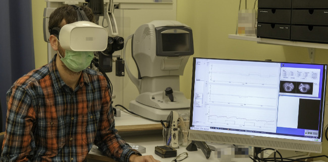
Purpose: The purpose of this study was to assess the feasibility of detecting relative afferent pupillary defects (RAPDs) using a commercial virtual reality headset equipped with an eye tracker.
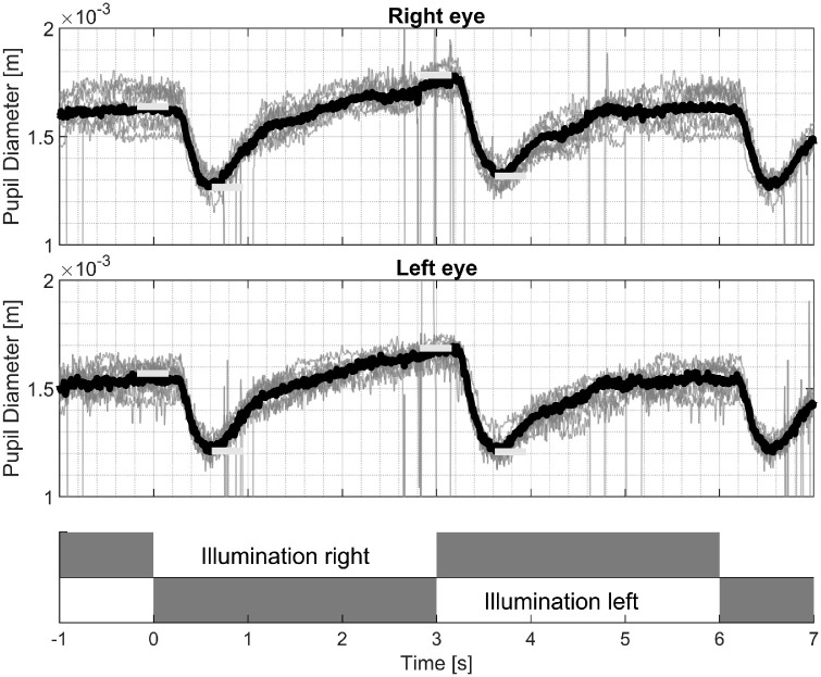
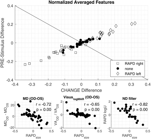
Methods: This is a cross-sectional study in which we compare the new computerized RAPD test with the traditional clinical standard using the swinging flashlight test. Eighty-two participants including 20 healthy volunteers aged 10 to 88 years were enrolled in this study. We present a bright/dark stimulus alternating between the eyes every 3 seconds using a virtual reality headset, and we simultaneously record changes in pupil size. To determine the presence of an RAPD, we developed an algorithm analyzing the pupil size differences. For the assessment of the performance of the automated and the manual measurement a post hoc impression based on all available data is created. The accuracy of the manual clinical evaluation and the computerized method is compared using confusion matrices and the gold standard of the post hoc impression. The latter is based on all available clinical information.
Results: We found that the computerized method detected RAPD with a sensitivity of 90.2% and an accuracy of 84.4%, as compared to the post hoc impression. This was not significantly different from the clinical evaluation with a sensitivity of 89.1% and an accuracy of 88.3%.
Conclusions: The presented method offers an accurate, easy to use, and fast method to measure an RAPD. In contrast to today's clinical practice, the measures are quantitative and objective.
Translational relevance: Computerized testing of Relative Afferent Pupillary Defects (RAPD) using a VR-headset and eye-tracking reaches non-inferior performance compared with senior neuro-ophthalmologists.
Conflict of interest statement
Disclosure:
Figures
References
-
- Relative Afferent Pupillary Defect - EyeWiki. Accessed September 9, 2022. Available at: https://eyewiki.aao.org/Relative_Afferent_Pupillary_Defect.
-
- Levatin P, Prasloski PF, Collen MF.. The swinging flashlight test in multiphasic screening for eye disease. Can J Ophthalmol. 1973; 8(2): 356–360. - PubMed
-
- Thompson HS. Afferent pupillary defects. Pupillary findings associated with defects of the afferent arm of the pupillary light reflex arc. Am J Ophthalmol. 1966; 62(5): 860–873. - PubMed
-
- Thompson HS, Corbett JJ, Cox TA.. How to measure the relative afferent pupillary defect. Survey Ophthalmol. 1981; 26(1): 39–42. - PubMed
-
- Boucher T, Fortin É, Evoy F.. The standard swinging flashlight test: reliable or not (P1.9-009). Neurology. 2019; 92(15 Supplement): P1.9–009.
