. 2023 Jun 19;13(6):764.
doi: 10.3390/metabo13060764.

Lactiplantibacillus plantarum and Saussurea costus as Therapeutic Agents against a Diabetic Rat Model-Approaches to Investigate Pharmacophore Modeling of Human IkB Kinase and Molecular Interaction with Dehydrocostus Lactone of Saussurea costus

Affiliations

Lactiplantibacillus plantarum and Saussurea costus as Therapeutic Agents against a Diabetic Rat Model-Approaches to Investigate Pharmacophore Modeling of Human IkB Kinase and Molecular Interaction with Dehydrocostus Lactone of Saussurea costus

Metab A AlGeffari et al. Metabolites. .

Abstract

Lactic acid bacteria is well-known as a vital strategy to alleviate or prevent diabetes. Similarly, the plant Saussurea costus (Falc) Lipsch is a preventive power against diabetes. Here, we aimed to determine whether lactic acid bacteria or Saussurea costus is more effective in treating a diabetic rat model in a comparative study manner. An in vivo experiment was conducted to test the therapeutic activity of Lactiplantibacillus plantarum (MW719476.1) and S. costus plants against an alloxan-induced diabetic rat model. Molecular, biochemical, and histological analyses were investigated to evaluate the therapeutic characteristics of different treatments. The high dose of S. costus revealed the best downregulated expression for the IKBKB, IKBKG, NfkB1, IL-17A, IL-6, IL-17F, IL-1β, TNF-α, TRAF6, and MAPK genes compared to Lactiplantibacillus plantarum and the control groups. The downregulation of IKBKB by S. costus could be attributed to dehydrocostus lactone as an active compound with proposed antidiabetic activity. So, we performed another pharmacophore modeling analysis to test the possible interaction between human IkB kinase beta protein and dehydrocostus lactone as an antidiabetic drug. Molecular docking and MD simulation data confirmed the interaction between human IkB kinase beta protein and dehydrocostus lactone as a possible drug. The target genes are important in regulating type 2 diabetes mellitus signaling, lipid and atherosclerosis signaling, NF-κB signaling, and IL-17 signaling pathways. In conclusion, the S. costus plant could be a promising source of novel therapeutic agents for treating diabetes and its complications. Dehydrocostus lactone caused the ameliorative effect of S. costus by its interaction with human IkB kinase beta protein. Further, future studies could be conducted to find the clinical efficacy of dehydrocostus lactone.

Keywords: 16S rRNA gene; Lactiplantibacillus plantarum; Saussurea costus; biochemical; diabetes mellitus; docking; histological analysis; pharmacophore modeling.


Conflict of interest statement

The authors declare no conflict of interest.

Figures

Figure 1
Figure 1
(A) In silico PCR of the current primer sequences matched with L. plantarum strains with an amplicon of 1251 bp, http://insilico.ehu.es/ accessed on 5 December 2022. (B) The created phylogenetic tree showed our current isolate with the nearest ones registered in the NCBI database.
Figure 2
Figure 2
LC MASS analysis showed the peak (231.5 m/z Da) of the chemical compound of dehydrocostus lactone, C15H18O2, from the extracted roots of S. costus.
Figure 3
Figure 3
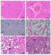
Micrograph of the H&E-stained sections of liver tissues of the (A) control group showing a normal histological structure of the liver with irregular plates of hepatocytes (H) radiating from the central vein (CV), separated by vascular sinusoids (S). (B) The control group shows the portal tract located at the peripheral part of the hepatic lobules, which includes a venule (a branch of the portal vein) (PV), an arteriole (a branch of the hepatic artery) (HA), and a bile ductule (BD). (C) A diabetic rat (alloxan-treated groups) demonstrates considerable disruption of hepatic architecture with generalized fatty change in hepatocytes, which gives the cytoplasm a foamy appearance. (D) A diabetic rat demonstrates most hepatocytes with peripheral nuclei; few hepatocytes show microvesicular steatosis (black circle). Notice the congested sinusoid (CS) and no clear lobular pattern. (E,F) A diabetic rat showing combined microvesicular (black circle) and macrovesicular (dotted arrows) fatty change, while a few hepatocytes appear normal (H), a few enlarged hepatocytes with peripheral flattened nuclei (thick black arrow), prominent periportal fibrous tissue (*) infiltrated with inflammatory cells (In). Notice the congested hepatic sinusoids (CS) and the congested blood vessels (CBV). (Stain: H&E; A, C: 100×, B, D, E, F: 400×).
Figure 4
Figure 4
Micrograph of the H&E-stained sections of the liver tissues of (A) diabetic rats treated with L. plantarum cell lysate showing hepatocytes with microvesicular (black circle) and macrovesicular steatosis (dotted arrow), normal hepatocytes (H), and inflammatory infiltration (steatohepatitis) (In). (B) The diabetic rats treated with L. plantarum intact cells showed normal morphology in most hepatocytes (H), a few hepatocytes showing large fat vacuoles (dotted arrow), as well as a few hepatocytes showing microvesicular fatty changes (black circle). Notice the congested hepatic sinusoids (CS) and the inflammatory infiltrate (In). (C) The diabetic rats treated with a low dose of S. costus show that most hepatocytes seem normal, some appear vacuolated with small steatotic vacuoles (black circle), and very few hepatocytes exhibit large steatotic vacuoles. Notice the inflammatory cell infiltration (In) between hepatocytes and the portal area’s connective tissue. (D) The diabetic rats treated with a high dosage of S. costus showed normal hepatic architecture; some hepatocytes exhibited microvesicular fatty change (black circle) and a few congested hepatic sinusoids (CS) (Stain: H&E; 100×).
Figure 5
Figure 5
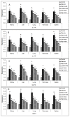
Impact of L. plantarum and S. costus on the regulation of (A) IKBKB, (B) IKBKG, (C) IL-1β, and (D) IL-6 genes in the liver, spleen, pancreas, lung, and kidney tissues of treated and non-treated rats. The β-actin gene was used to normalize the comparative mRNA levels. a,b,c,d,e,f: bars with different letters in the same tissue differ significantly (p < 0.05).
Figure 6
Figure 6
Impact of L. plantarum and S. costus on regulating (A) IL-17A, (B) IL-17F, (C) Mitogen-activated protein kinase 1 (MAPK), and (D) NFKB1 genes in the liver, spleen, pancreas, lung, and kidney tissues of treated and non-treated rats. The β-actin gene was used to normalize the comparative mRNA levels. a,b,c,d,e,f: bars with different letters in the same tissue differ significantly (p < 0.05).
Figure 7
Figure 7
The impact of L. plantarum and S. costus on (A) TNF-α and (B) TRAF6 gene regulation in the liver, spleen, pancreas, lung, and kidney tissues of treated and non-treated rats. The β-actin gene was used to normalize the comparative mRNA levels. a,b,c,d,e,f: means with different letters in the same tissue differ significantly (p < 0.05).
Figure 8
Figure 8
The 3D chemical structure of the selected ligands for molecular docking (dehydrocostus lactone, bigelovin, caprolactone, and methyl nonanoate ester. SMILES were retrieved from the PubChem database https://pubchem.ncbi.nlm.nih.gov accessed on 14 May 2023 and were drawn with the Marvin JS tool https://marvinjs-demo.chemaxon.com/latest/demo.html accessed on 14 May 2023.
Figure 9
Figure 9
4KIK protein showed the pocket of ligand dehydrocostus lactone after docking using UCSF-Chimera version 1.17.1 and visualized it (A,B). The obtained affinity score was (−8.619 kcal/mol). The ligand–protein interaction was performed in Maestro software version 2022-4 and Discovery Studio version 2021 (C,D).
Figure 10
Figure 10
4KIK protein showed the pocket of ligand bigelovin after docking using UCSF-Chimera version 1.17.1 and visualized it (A,B). The obtained affinity score was (−7.882 kcal/mol). The ligand–protein interaction was performed in Maestro version 2022-4 and Discovery Studio version 2021 (C,D).
Figure 11
Figure 11
4KIK protein showed the pocket of caprolactone after docking using UCSF-Chimera version 1.17.1 and visualized it (A,B). The obtained affinity score was (−5.574 kcal/mol). The ligand–protein interaction was performed in Maestro version 2022-4 and Discovery Studio version 2021 (C,D).
Figure 12
Figure 12
4KIK protein showed the pocket of the methyl nonanoate ester after docking using UCSF-Chimera version 1.17.1 and visualized it (A,B). The obtained affinity score was (−4.997 kcal/mol). The ligand–protein interaction was performed in Maestro version 2022-4 and Discovery Studio version 2021(C,D).
Figure 13
Figure 13
(A) The three proteins (4KIK, 4WSQ, and 5M5A) and (B) five proteins (4KIK, 4WSQ, 5M5A, 3EQF, and 1ROP) kinases were compared with the same ligand KSA (448239) in MatchMaker, UCSF-Chimera version 1.17.1.
Figure 14
Figure 14
GROMACS energies chart displayed temperature with a black curve, while T-Protein-LIG was a red color curve. (A) RMS fluctuation chart displayed proteins with a black curve, a backbone with a red curve, and ligands with a green curve (B). The radius of gyration (total and around axes) chart showed proteins with a black curve, a backbone with a red curve, and a ligand with a green curve, respectively (C). The previous GROMACS analysis was performed on GROMACS version 2022.4 for 4KIK protein with dehydrocostus lactone and was visualized with Grace version 5.1.25.
Figure 15
Figure 15
RMSD chart showed ligand–protein with a black curve, ligand–backbone with a red curve, and Na–protein with a green curve, respectively (A). The hydrogen bonds chart displayed protein with ligands, Na, and ions at the same level in green (B). The previous GROMACS analysis was performed on GROMACS version 2022.4 for 4KIK protein with dehydrocostus lactone and was visualized with Grace version 5.1.25.

Similar articles

Cited by

References

    1. Lim L.L., Chow E., Chan J.C.N. Cardiorenal Diseases in Type 2 Diabetes Mellitus: Clinical Trials and Real-World Practice. Nat. Rev. Endocrinol. 2023;19:151–163. doi: 10.1038/s41574-022-00776-2. - DOI - PubMed
    1. Davies M.J., D’Alessio D.A., Fradkin J., Kernan W.N., Mathieu C., Mingrone G., Buse J.B. Management of Hyperglycaemia in Type 2 Diabetes. A Consensus Report by the American Diabetes Association (ADA) and the European Association for the Study of Diabetes (EASD) Diabetologia. 2022;65:1925–1966. doi: 10.1007/s00125-022-05787-2. - DOI - PMC - PubMed
    1. Hegde P.K., Rao H.A., Rao P.N. A Review on Insulin Plant (Costus igneus Nak) Pharmacogn. Rev. 2014;8:67–72. - PMC - PubMed
    1. Mammate N., El Oumari F.E., Imtara H., Belchkar S., Lahrichi A., Alqahtani A.S., Houssaini T.S. Antioxidant and Anti-Urolithiatic Activity of Aqueous and Ethanolic Extracts from Saussurea costus (Falc) Lispich Using Scanning Electron Microscopy. Life. 2022;12:1026. doi: 10.3390/life12071026. - DOI - PMC - PubMed
    1. Kumar A., Sharma B. Biomedical implications of plant-based principles as antidepressants: Prospects for novel drug development. Mini Rev. Med. Chem. 2022;22:904–926. - PubMed

LinkOut - more resources