. 2023 Aug 8;8(15):e170121.
doi: 10.1172/jci.insight.170121.

An LRP1-binding motif in cellular prion protein replicates cell-signaling activities of the full-length protein

An LRP1-binding motif in cellular prion protein replicates cell-signaling activities of the full-length protein

Elisabetta Mantuano et al. JCI Insight. .

Abstract

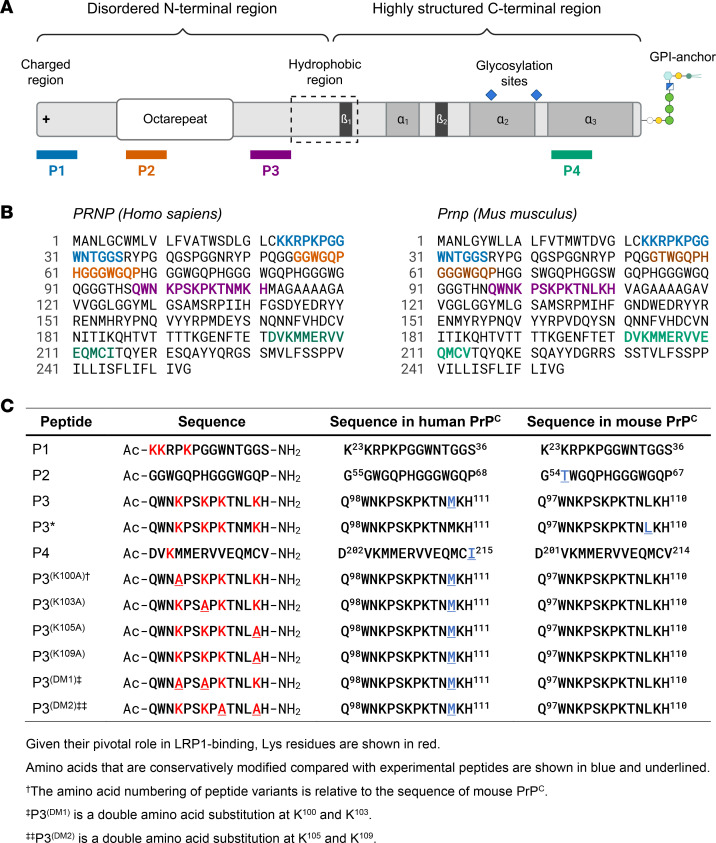
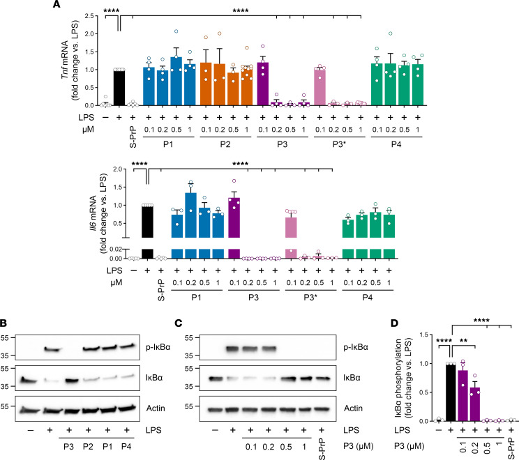
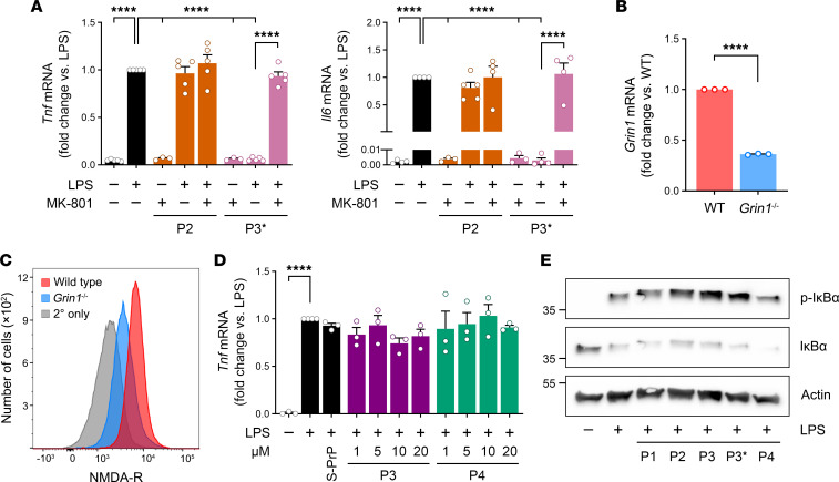
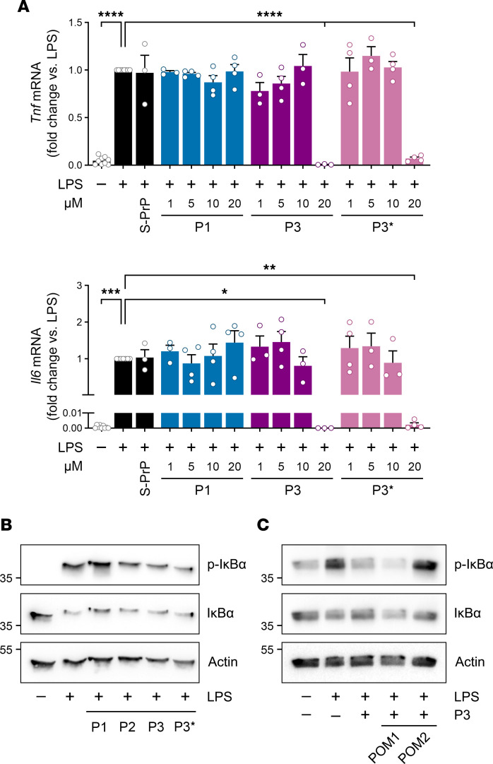
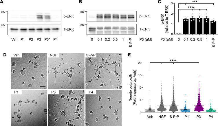
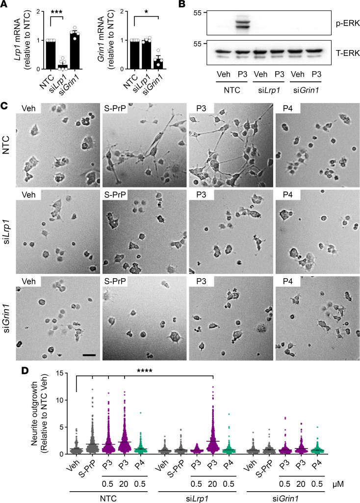
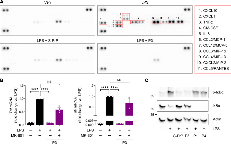
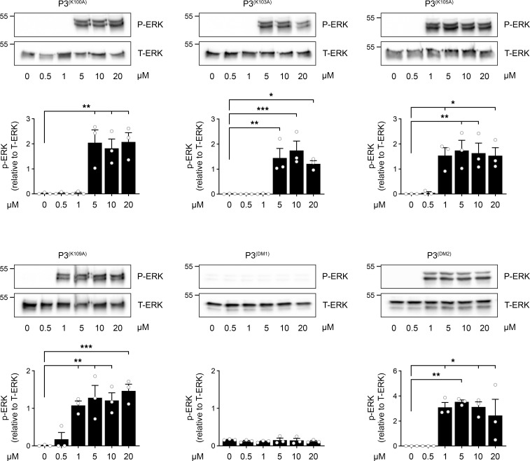
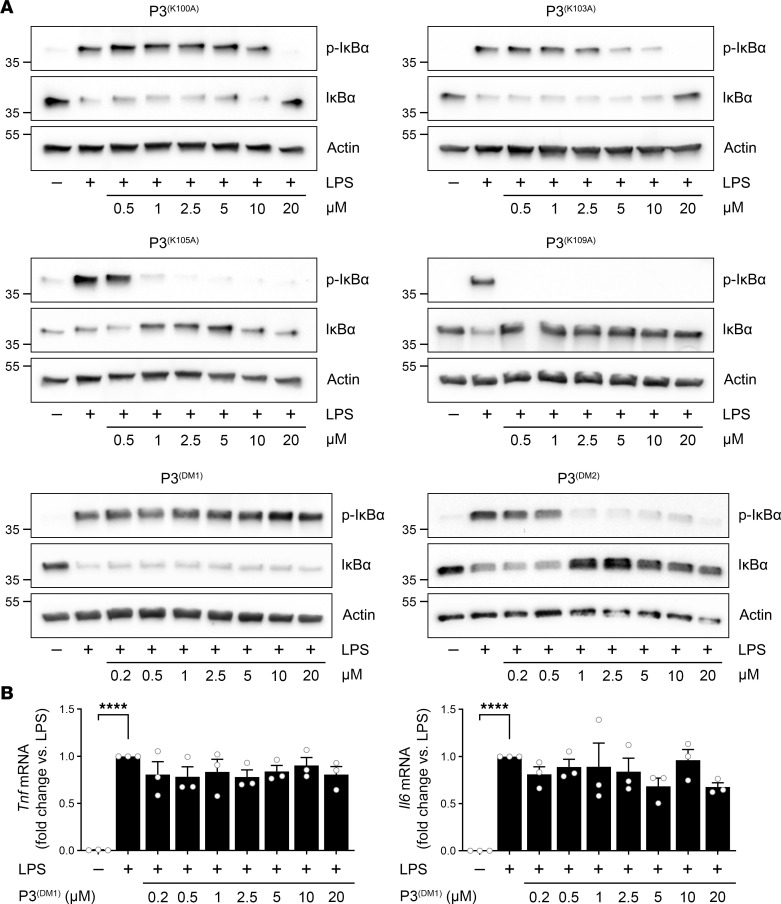
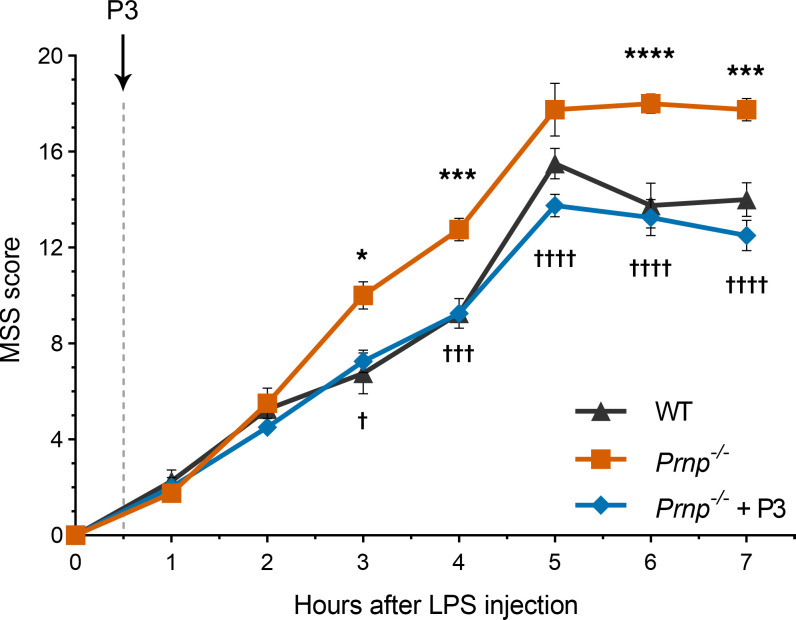
Low-density lipoprotein receptor-related protein-1 (LRP1) functions as a receptor for nonpathogenic cellular prion protein (PrPC), which is released from cells by ADAM (a disintegrin and metalloproteinase domain) proteases or in extracellular vesicles. This interaction activates cell signaling and attenuates inflammatory responses. We screened 14-mer PrPC-derived peptides and identified a putative LRP1 recognition motif in the PrPC sequence spanning residues 98-111. A synthetic peptide (P3) corresponding to this region replicated the cell-signaling and biological activities of full-length shed PrPC. P3 blocked LPS-elicited cytokine expression in macrophages and microglia and rescued the heightened sensitivity to LPS in mice in which the PrPC gene (Prnp) had been deleted. P3 activated ERK1/2 and induced neurite outgrowth in PC12 cells. The response to P3 required LRP1 and the NMDA receptor and was blocked by the PrPC-specific antibody, POM2. P3 has Lys residues, which are typically necessary for LRP1 binding. Converting Lys100 and Lys103 into Ala eliminated the activity of P3, suggesting that these residues are essential in the LRP1-binding motif. A P3 derivative in which Lys105 and Lys109 were converted into Ala retained activity. We conclude that the biological activities of shed PrPC, attributed to interaction with LRP1, are retained in synthetic peptides, which may be templates for therapeutics development.

Keywords: Cell Biology; Inflammation; Peptides; Prions; Signal transduction.


Conflict of interest statement

Conflict of interest: The authors have declared that no conflict of interest exists.

Figures

Figure 1
Figure 1. Synthetic peptides and their relation to the structure of PrPC.
(A) Location of the primary set of 4 synthetic peptides in relation to the structure of PrPC. (B) Using the same color-coding system applied in A, P1–P4 are located within the primary sequences of human and mouse PrPC. (C) The sequences of all studied synthetic peptides, including variants of P3/P3*, are shown. Lys residues and Lys residues that were converted to Ala in second-generation peptides are shown in red. Conservative sequence differences between the synthetic peptides and the structure of human and mouse PrPC are shown in blue and underlined.
Figure 2
Figure 2. P3 replicates the effects of S-PrP and EV-associated PrPC in macrophages.
(A) BMDMs from C57BL/6J mice were treated for 6 hours with LPS (0.1 μg/mL) in the presence or absence of S-PrP (40 nM) or increasing concentrations (0.1–1.0 μM) of P1, P2, P3, P3*, or P4. RT-qPCR was performed to determine mRNA levels of Tnf and Il6 (mean ± SEM, n = 3–9, individual points shown; 1-way ANOVA: ****P < 0.0001). (B) BMDMs were treated for 1 hour with LPS (0.1 μg/mL) in the presence or absence of P1, P2, P3, or P4 (each at 0.5 μM). Immunoblot analysis was performed to detect phosphorylated IκBα, total IκBα, and β-actin. (C) BMDMs were treated for 1 hour with LPS (0.1 μg/mL) in the presence or absence of S-PrP (40 nM) or increasing concentrations of P3 (0.1–1 μM). Immunoblot analysis was performed to detect phosphorylated IκBα, total IκBα, and β-actin. (D) Densitometry analysis of phosphorylated IκBα band intensity relative to β-actin for cells treated with LPS and different concentrations of P3 (mean ± SEM, n = 3, 1-way ANOVA: **P < 0.01, ****P < 0.0001).
Figure 3
Figure 3. The NMDA-R is necessary for the response to P3 in macrophages.
(A) BMDMs were pretreated with MK-801 (1 μM) or vehicle for 30 minutes. The cells were then treated with LPS (0.1 μg/mL), P2 (0.5 μM), or P3* (0.5 μM), for 6 hours, as indicated. RT-qPCR was performed to compare mRNA levels for Tnf and Il6 (mean ± SEM, n = 3–7, individual points are shown; 1-way ANOVA: ****P < 0.0001). (B) BMDMs were harvested from Grin1fl/fl LysM-Cre+ mice. Grin1 mRNA expression was determined by RT-qPCR and compared with that detected in BMDMs isolated from Grin1fl/fl LysM-Cre mice (n = 3; mean ± SEM; unpaired 2-tailed t test: ****P < 0.0001). (C) Flow cytometry was performed to detect cell surface GluN1 NMDA-R subunit in BMDMs isolated from Grin1fl/fl LysM-Cre–positive and –negative (wild-type) mice. As a control, cells from LysM-Cre mice were incubated with secondary antibody only (gray). (D) BMDMs from Grin1fl/fl LysM-Cre+ mice were treated for 6 hours with LPS (0.1 μg/mL), in the presence of S-PrP (40 nM) or increasing concentrations of P3 (1–20 μM), P4 (1–20 μM), or vehicle. RT-qPCR was performed to determine Tnf mRNA (mean ± SEM, n = 3; 1-way ANOVA: ****P < 0.0001). (E) BMDMs from Grin1fl/fl LysM-Cre+ mice were treated for 1 hour with LPS (0.1 μg/mL), in the presence of P1, P2, P3, P3*, and P4, as indicated (each at 0.5 μM). Immunoblot analysis was performed to detect phosphorylated IκBα, total IκBα, and β-actin.
Figure 4
Figure 4. The antiinflammatory activity of P3/P3* is strongly facilitated by LRP1 and blocked by POM2.
(A) BMDMs from Lrp1fl/fl LysM-Cre+ mice were treated for 6 hours with LPS (0.1 μg/mL) in the presence of S-PrP (40 nM) or increasing concentrations (1–20 μM) of P1, P3, P3*, or vehicle. RT-qPCR was performed to determine mRNA levels for Tnf and Il6 (mean ± SEM, n = 3–4; 1-way ANOVA: *P < 0.05, **P < 0.01, ***P < 0.001, ****P < 0.0001). (B) BMDMs were treated for 1 hour with LPS (0.1 μg/mL) in the presence of P1, P2, P3, or P3* (each at 0.5 μM). Immunoblot analysis was performed to detect p-IκBα, total IκBα, and β-actin. (C) BMDMs were treated for 1 hour with LPS (0.1 μg/mL) and P3 (0.5 μM), in the presence of POM1 or POM2 (10 μg/mL), as indicated. Immunoblot analysis was performed to detect p-IκBα, total IκBα, and β-actin.
Figure 5
Figure 5. P3 activates cell signaling and promotes neurite outgrowth in PC12 cells.
(A) PC12 cells were treated with P1, P2, P3, P3*, and P4 (each at 0.5 μM) for 10 minutes. Cell extracts were subjected to immunoblot analysis to detect p-ERK1/2 and total ERK1/2. (B) PC12 cells were stimulated for 10 minutes with increasing concentrations of P3 (0.1–1.0 μM) or with S-PrP (40 nM). Phosphorylated ERK1/2 and total ERK1/2 were determined. (C) Densitometry analysis of p-ERK1/2 relative to total ERK1/2 (T-ERK) in PC12 cells treated with P3 or S-PrP. The bars represent the mean ± SEM of the results from 3 separate experiments (1-way ANOVA: ***P < 0.001, ****P < 0.0001). (D) PC12 cells were treated for 48 hours with S-PrP (40 nM), P1 (0.5 μM), P3 (0.5 μM), P4 (0.5 μM), NGF-β (50 ng/mL) as a positive control, or vehicle. Neurite outgrowth was examined by phase contrast microscopy. Representative images are shown (scale bar, 50 μm). (E) Neurite length was determined by analyzing all the cells in ≥5 random fields per treatment, in 3 different experiments (mean ± SEM; 1-way ANOVA: ****P < 0.0001).
Figure 6
Figure 6. P3 activates ERK1/2 and promotes neurite outgrowth in PC12 cells by a mechanism that requires the NMDA-R and LRP1.
(A) PC12 cells were transfected with siRNA specifically targeting Lrp1 or Grin1. Control cells were transfected with NTC siRNA. Expression of the mRNAs encoding LRP1 and the GluN1 NMDA-R subunit was determined 48 hours later by RT-qPCR (n = 4–6; mean ± SEM; 1-way ANOVA: *P < 0.05; ***P < 0.001). (B) PC12 cells were transfected with Lrp1-specific siRNA, Grin1-specific siRNA, or NTC siRNA and then treated with P3 (0.5 μM) or vehicle for 10 minutes. ERK1/2 activation (p-ERK) was determined by immunoblotting. (C) PC12 cells were transfected with Lrp1-specific siRNA, Grin1-specific siRNA, or NTC siRNA, as indicated. The cells were then treated with S-PrP (40 nM), P3 (0.5 μM), or P4 (0.5 μM) for 48 hours. Neurite outgrowth was detected by phase contrast microscopy. Representative images are shown (scale bar, 50 μm). (D) Results are summarized for the studies shown in C and for PC12 cells treated with 20 μM P3. Neurite length was determined in all the cells of ≥5 random fields per treatment, in 3 different experiments (mean ± SEM; 1-way ANOVA: ****P < 0.0001).
Figure 7
Figure 7. P3 inhibits the pro-inflammatory activity of LPS in microglia.
(A) Microglia were isolated from C57BL/6J mouse pups and treated with LPS (0.1 μg/mL) for 6 hours, in the presence and absence of S-PrP (40 nM) or P3 (0.5 μM). Conditioned medium (CM) was collected and analyzed using Proteome Profiler Mouse Cytokine Array Kit (R&D Systems). Representative cytokines that were increased in CM when LPS was added in the absence of S-PrP or P3 are numbered in red boxes. (B) Microglia were treated with LPS (0.1 μg/mL) P3 (0.5 μM), and MK-801 (1 μM), as indicated. RT-qPCR was performed to determine mRNA levels of Tnf and Il6 (mean ± SEM; n = 3; 1-way ANOVA: ****P < 0.0001). (C) Microglia were treated for 1 hour with LPS (0.1 μg/mL) in the presence or absence of S-PrP (40 nM), P1, P3, or P4 (0.5 μM). Immunoblot analysis was performed to detect p-IκBα, total IκBα, and β-actin.
Figure 8
Figure 8. Lys100 and Lys103 are required for the function of P3 in PC12 cells.
PC12 cells were treated for 10 minutes with increasing concentrations (0.5–20 μM) of P3(K100A), P3(K103A), P3(K105A), P3(K109A), P3(DM1), or P3(DM2). Immunoblot analysis was performed to determine ERK1/2 phosphorylation. Densitometry analysis shows p-ERK1/2 relative to total ERK1/2 (T-ERK). The bars represent the mean ± SEM of the results from 3 separate experiments (1-way ANOVA: *P < 0.05; **P < 0.01; ***P < 0.001).
Figure 9
Figure 9. Lys100 and Lys103 are required for the function of P3 in macrophages.
(A) BMDMs from wild-type mice were treated for 1 hour with LPS (0.1 μg/mL) and increasing concentrations (0.2–20 μM) of P3(K100A), P3(K103A), P3(K105A), P3(K109A), P3(DM1), or P3(DM2), as indicated above each panel. Immunoblot analysis was performed to detect p-IκBα, total IκBα, and β-actin. (B) BMDMs from wild-type mice were treated for 6 hours with LPS (0.1 μg/mL) in the presence of increasing concentrations of P3(DM1) (0.2–20 μM). RT-qPCR was performed to determine mRNA levels for Tnf and Il6 (mean ± SEM; n = 3; 1-way ANOVA: ****P < 0.0001).
Figure 10
Figure 10. P3 rescues the increased susceptibility of Prnp–/– mice to LPS.
Male 16- to 20-week old Prnp–/– mice (shown in orange) and wild-type mice in the same genetic background (shown in black) were challenged with LPS, by IP injection, at 75% of the LD50. A second matched cohort of Prnp–/– mice was treated with LPS and then with P3, 0.5 hour later (blue). Toxicity was scored as described in Methods. Prnp–/– mice demonstrated significantly more toxicity compared with wild-type mice (mean ± SEM; n = 4; 2-way ANOVA: *P < 0.05; ***P < 0.001; ****P < 0.0001). P3 significantly reversed the toxicity of LPS in Prnp–/– mice (mean ± SEM; n = 4; 2-way ANOVA: P < 0.05; †††P < 0.001; ††††P < 0.0001). MSS, mouse sepsis score.

Similar articles

Cited by

References

    1. Herz J, Strickland DK. LRP: a multifunctional scavenger and signaling receptor. J Clin Invest. 2001;108(6):779–784. doi: 10.1172/JCI200113992. - DOI - PMC - PubMed
    1. Fernandez-Castaneda A, et al. Identification of the low density lipoprotein (LDL) receptor-related protein-1 interactome in central nervous system myelin suggests a role in the clearance of necrotic cell debris. J Biol Chem. 2013;288(7):4538–4548. doi: 10.1074/jbc.M112.384693. - DOI - PMC - PubMed
    1. Basu S, et al. CD91 is a common receptor for heat shock proteins gp96, hsp90, hsp70, and calreticulin. Immunity. 2001;14(3):303–313. doi: 10.1016/S1074-7613(01)00111-X. - DOI - PubMed
    1. Rauch JN, et al. LRP1 is a master regulator of tau uptake and spread. Nature. 2020;580(7803):381–385. doi: 10.1038/s41586-020-2156-5. - DOI - PMC - PubMed
    1. Chen K, et al. LRP1 is a neuronal receptor for α-synuclein uptake and spread. Mol Neurodegener. 2022;17(1):57. doi: 10.1186/s13024-022-00560-w. - DOI - PMC - PubMed

Publication types