. 2023 Jun 13;9(3):1137-1152.
doi: 10.3390/tomography9030094.

Sinogram Inpainting with Generative Adversarial Networks and Shape Priors

Affiliations

Sinogram Inpainting with Generative Adversarial Networks and Shape Priors

Emilien Valat et al. Tomography. .

Abstract

X-ray computed tomography is a widely used, non-destructive imaging technique that computes cross-sectional images of an object from a set of X-ray absorption profiles (the so-called sinogram). The computation of the image from the sinogram is an ill-posed inverse problem, which becomes underdetermined when we are only able to collect insufficiently many X-ray measurements. We are here interested in solving X-ray tomography image reconstruction problems where we are unable to scan the object from all directions, but where we have prior information about the object's shape. We thus propose a method that reduces image artefacts due to limited tomographic measurements by inferring missing measurements using shape priors. Our method uses a Generative Adversarial Network that combines limited acquisition data and shape information. While most existing methods focus on evenly spaced missing scanning angles, we propose an approach that infers a substantial number of consecutive missing acquisitions. We show that our method consistently improves image quality compared to images reconstructed using the previous state-of-the-art sinogram-inpainting techniques. In particular, we demonstrate a 7 dB Peak Signal-to-Noise Ratio improvement compared to other methods.

Keywords: Generative Adversarial Network; X-ray computed tomography; computer assisted design data; machine-learning.


Conflict of interest statement

The authors declare no conflict of interest.

Figures

Figure 1
Figure 1
The pix2pix architecture using a generator and a discriminator. The generator is trained to generate missing sinogram information from a multi-channel input. One channel is the sinogram with the missing information, one is a sinogram encoding the object shape and one is a mask indicating where the CAD expects missing information to be located. The discriminator (or detector) is a classification network that tries to detect whether the multi-channel input is an actual full sinogram or is a sinogram where the generator estimates missing information. GAN training is done iteratively, with the generator trying to fool the discriminator better and the discriminator trying to improve the detection of estimated data.
Figure 2
Figure 2
Sample of the input data given to the generator. We estimate the full sinogram (a) from the sinogram with missing data (b), the sinogram of the shape prior (c) and the sinogram’s mask indicating the location of missing projections (d).
Figure 3
Figure 3
Explanation of the training procedure. The CAD sinogram is added to both target and limited-view sinograms.
Figure 4
Figure 4
Real training samples. A 2D slice from the SophiaBeads dataset, showing the full slice we want to estimate (a) and the image that encodes the shape prior (b). Here the shape prior indicates each of the locations for each sphere in the image. However, as the attenuation value of each sphere is unknown, random attenuation values are assigned.
Figure 5
Figure 5
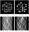
Slice samples from the synthetic dataset. The ground-truth reconstruction (a), and the shape prior (b) showing each object with a different randomly assigned grey value, in which no plastic container is visible. In an attempt to reproduce noise in the image, Gaussian noise is added to the material of uniform densities. (b) Sinogram samples from the synthetic dataset, from the ground-truth reconstruction (c) and from the shape prior (d). The observed sinograms are obtained using the forward radon transform with a parallel beam geometry.
Figure 6
Figure 6
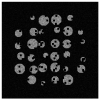
Simulated XCT images with objects with internal defects that are not encoded in shape prior.
Figure 7
Figure 7
Reconstructions of a slice from one of the SophiaBeads test datasets. First, the target image (a), reconstructed from 256 measurements. Then, the image reconstructed from half of the measurements (b), the linear interpolation (c), the replacement of the missing measurements by the ones expected by the CAD (d), the measurements inferred by the Unet method without CAD data (e), the measurements inferred by the Unet method with CAD data (f), the measurements inferred by the GAN without CAD data (g) and the measurements inferred by our method (h).
Figure 7
Figure 7
Reconstructions of a slice from one of the SophiaBeads test datasets. First, the target image (a), reconstructed from 256 measurements. Then, the image reconstructed from half of the measurements (b), the linear interpolation (c), the replacement of the missing measurements by the ones expected by the CAD (d), the measurements inferred by the Unet method without CAD data (e), the measurements inferred by the Unet method with CAD data (f), the measurements inferred by the GAN without CAD data (g) and the measurements inferred by our method (h).
Figure 8
Figure 8
Reconstructions of the synthetic samples using various methods. First, the one from the sinogram 256 measurements (a), then from the sinogram interpolated with our method (b), the difference between the latter and the target (c), the one interpolated with the shape prior (d) and the difference between the latter and the target (e). When reconstructing data with defects using shape priors, the method can identify the objects’ location, but the defect’s fine details are more difficult to identify.

References

    1. Hall E.J., Brenner D.J. Cancer risks from diagnostic radiology. Br. J. Radiol. 2008;81:362–378. doi: 10.1259/bjr/01948454. - DOI - PubMed
    1. du Plessis A., le Roux S.G., Guelpa A. Comparison of medical and industrial X-ray computed tomography for non-destructive testing. Case Stud. Nondestruct. Test. Eval. 2016;6:17–25. doi: 10.1016/j.csndt.2016.07.001. - DOI
    1. Feldkamp L.A., Davis L.C., Kress J.W. Practical Cone-Beam Algorithm. Ford Motor Company; Dearborn, MI, USA: 1984. Technical Report 6.
    1. Biguri A., Dosanjh M., Hancock S., Soleimani M. TIGRE: A MATLAB-GPU toolbox for CBCT image reconstruction. Biomed. Phys. Eng. Express. 2016;2:055010. doi: 10.1088/2057-1976/2/5/055010. - DOI
    1. Candès E.J., Romberg J., Tao T. Robust uncertainty principles: Exact signal reconstruction from highly incomplete frequency information. IEEE Trans. Inf. Theory. 2006;52:489–509. doi: 10.1109/TIT.2005.862083. - DOI

Publication types

MeSH terms