. 2023 Jun 28;8(1):247.
doi: 10.1038/s41392-023-01453-0.

Extracellular matrix-derived mechanical force governs breast cancer cell stemness and quiescence transition through integrin-DDR signaling

Affiliations

Extracellular matrix-derived mechanical force governs breast cancer cell stemness and quiescence transition through integrin-DDR signaling

Cong Li et al. Signal Transduct Target Ther. .

Abstract

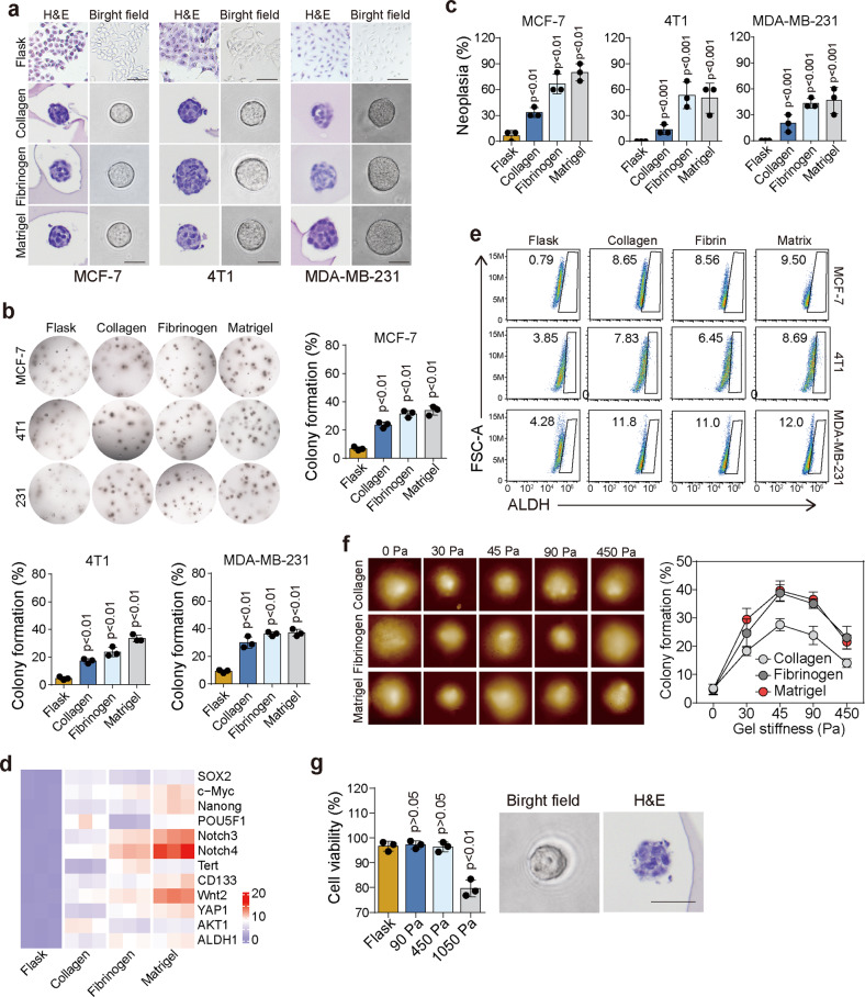
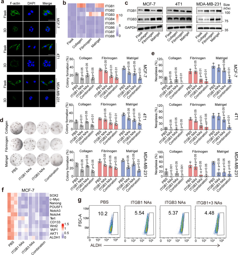
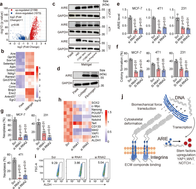
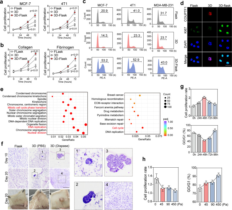
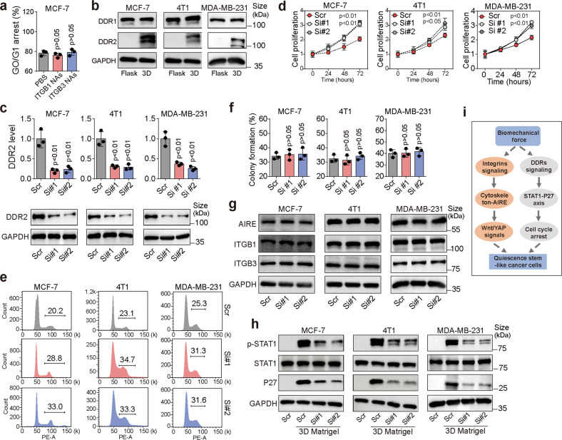
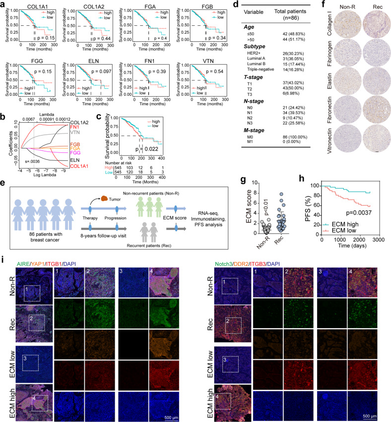
The extracellular matrix (ECM) serves as signals that regulate specific cell states in tumor tissues. Increasing evidence suggests that extracellular biomechanical force signals are critical in tumor progression. In this study, we aimed to explore the influence of ECM-derived biomechanical force on breast cancer cell status. Experiments were conducted using 3D collagen, fibrinogen, and Matrigel matrices to investigate the role of mechanical force in tumor development. Integrin-cytoskeleton-AIRE and DDR-STAT signals were examined using RNA sequencing and western blotting. Data from 1358 patients and 86 clinical specimens were used for ECM signature-prognosis analysis. Our findings revealed that ECM-derived mechanical force regulated tumor stemness and cell quiescence in breast cancer cells. A mechanical force of ~45 Pa derived from the extracellular substrate activated integrin β1/3 receptors, stimulating stem cell signaling pathways through the cytoskeleton/AIRE axis and promoting tumorigenic potential and stem-like phenotypes. However, excessive mechanical force (450 Pa) could drive stem-like cancer cells into a quiescent state, with the removal of mechanical forces leading to vigorous proliferation in quiescent cancer stem cells. Mechanical force facilitated cell cycle arrest to induce quiescence, dependent on DDR2/STAT1/P27 signaling. Therefore, ECM-derived mechanical force governs breast cancer cell status and proliferative characteristics through stiffness alterations. We further established an ECM signature based on the fibrinogen/fibronectin/vitronectin/elastin axis, which efficiently predicts patient prognosis in breast cancer. Our findings highlight the vital role of ECM-derived mechanical force in governing breast cancer cell stemness/quiescence transition and suggest the novel use of ECM signature in predicting the clinical prognosis of breast cancer.


Conflict of interest statement

The authors declare no competing interests.

Figures

Fig. 1
Fig. 1
The ECM-induced biomechanical force promoted breast tumor stemness. a Representative images and H&E staining images of MCF-7, 4T1 and MDA-MB-231 cells seeded in a flask system and different 3D gels (collagen, fibrinogen, and Matrigel) for 3 days. The scale bar was 50 μm. b, c MCF-7, 4T1 and MDA-MB-231 cells were cultured in a flask system or 3D gels (collagen, fibrinogen, and Matrigel) for 3 days. In vitro colony formation (b) and in vivo tumor formation assay (n = 10) (c) were performed. d Heatmap of stemness-associated genes (SOX2, c-Myc, Nanog, POU5F1, Notch3, Notch4, Tert, CD133, Wnt2, YAP1, AKT1, and ALDH1) expression in MCF-7 cells cultured in flask and different 3D gels (collagen, fibrinogen, and Matrigel) for 3 days, determined using qPCR. e MCF-7, 4T1 and MDA-MB-231 cells were cultured in a flask system or 3D gels (collagen, fibrinogen, and Matrigel) for 3 days. ALDH1+ cell subpopulations were determined by flow cytometry. f MCF-7 cells were seeded in different 3D gels (collagen, fibrinogen, and Matrigel) with different stiffness (0, 30, 45, 90, and 450 Pa) for 3 days. Following this, the in vitro colony formation assay was performed. Representative images of tumor cells during atomic force microscopy analysis are shown. g Viability of MCF-7 cells seeded in a flask or 3D Matrigel (90, 450, and 1050 Pa). Representative image and H&E staining of MCF-7 cells seeded in 3D Matrigel (1050 Pa, 3 days) are shown. The scale bar is 50 μm. Three independent experiments were performed. Data are represented as mean ± SEM. P < 0.05, statistical significance
Fig. 2
Fig. 2
ECM compounds bind to integrins to transduce biomechanical force signals. a Immunostaining of F-actin in the MCF-7, 4T1 and MDA-MB-231 cells cultured in a flask and 3D Matrigel for 3 days. The scale bar is 20 μm. b Heatmap of integrin β1 ~ 8 expression in MCF-7 cells cultured in a flask and different 3D gels (collagen, fibrinogen, and Matrigel) for 3 days. c Western blotting of integrin β1 and β3 in MCF-7, 4T1 and MDA-MB-231 cells cultured in a flask and different 3D gels (collagen, fibrinogen, and Matrigel) for 3 days. d, e in vitro colony formation (d) and in vivo tumor formation (n = 10) (e) assays for MCF-7/4T1/MDA-MB-231 cells seeded in different 3D gels (collagen, fibrinogen, and Matrigel) and treated with PBS and integrin β1- and β3-neutralizing antibodies, respectively. f Heatmap of stemness-associated gene (SOX2, c-Myc, Nanog, POU5F1, Notch3, Notch4, Tert, CD133, Wnt2, YAP1, AKT1, and ALDH1) expression in MCF-7 cells (3D Matrigel culture) treated with PBS and integrin β1- and β3-neutralizing antibodies. g ALDH1+ cell subpopulations were determined in MCF-7 cells (3D Matrigel culture) treated with PBS and integrin β1- and β3-neutralizing antibodies. Three independent experiments were performed. Data are represented as mean ± SEM. P < 0.05, statistical significance
Fig. 3
Fig. 3
Integrin-cytoskeleton-AIRE signals are crucial for stemness gene upregulation. a Volcano plots showing the differentially expressed genes in 4T1 cells cultured in a flask and 3D Matrigel for 3 days. b Heatmap of top 15 upregulated genes in 3D Matrigel-cultured 4T1 cells in comparison with that of cells cultured in a flask. c Western blotting for AIRE in MCF-7, 4T1 and MDA-MB-231 cells cultured in a flask or 3D Matrigel (treated with PBS, integrin β1/3-neutralizing antibodies, or 5a-Pregnane-3,20-dione). d Western blotting for AIRE in MCF-7 cells cultured in a flask or 3D collagen/fibrinogen gels for 3 days. e AIRE expression at the mRNA level in 3D Matrigel-cultured MCF-7/4T1/MDA-MB-231 cells, treated with scramble or AIRE siRNA. f, g in vitro colony formation potential (f) and in vivo tumor formation (n = 10)(g) potential of 3D Matrigel-cultured MCF-7/4T1/MDA-MB-231 cells treated with scramble or AIRE siRNA. h Heatmap of stemness-associated gene (SOX2, c-Myc, Nanog, POU5F1, Notch3, Notc4, Tert, CD133, Wnt2, YAP1, AKT1, and ALDH1) expression in 3D Matrigel cultured MCF-7 cells treated with scramble or AIRE siRNA. i ALDH1+ cell subpopulations were determined in MCF-7 cells (3D Matrigel culture) treated with scramble or AIRE siRNA. j Schematic representation of the integrin-cytoskeleton-AIRE signals in breast cancer cells. Three independent experiments were performed. Data are represented as mean ± SEM. P < 0.05, statistical significance
Fig. 4
Fig. 4
ECM-induced biomechanical force drives stem cell-like tumor cell quiescence. a MCF-7/4T1 cells were seeded in a flask and 3D Matrigel, following which the cells were harvested for the cell proliferation assay in a 96-well plate (flask and 3D-flask groups). Some of the 3D-cultured MCF-7/4T1 cells were re-seeded in 3D Matrigel and subjected to cell proliferation determination at the same time points (3D group). b Proliferation of MCF-7 cells cultured in a flask, 3D-flask, and 3D gel (3D collagen and fibrinogen culture system). c Cell cycle analysis of MCF-7/4T1/MDA-MB-231 cells cultured in a flask, 3D gels, and 3D-flask. d Immunostaining for Ki67 and CoupTF1 in the flask, 3D gel, and 3D-flask groups. The scale bar is 20 μm. e GO and KEGG enrichment analysis of differentially expressed genes in the 4T1 cells cultured in the flask and 3D culture system, with a significance threshold of p-value < 0.05. f 1 × 103 4T1 cells were encapsulated in a 450-Pa 3D Matrigel (or not) and subcutaneously implanted into mice. On days 3 and 5, the mice were treated with PBS or dispase administered via subcutaneous injection. H&E staining of the hypodermis in each group was performed on days 10 and 20 (n = 10). The scale bar was 500 μm. g MCF-7 cells were cultured in the 3D Matrigel for 3 days and isolated for flask culture. After 0, 24, 48, 72, and 96 h of flask culture, cell proliferation or cycle was examined. h MCF-7 cells were seeded in 45-, 90-, and 450-Pa Matrigel for 3 days. Cell cycle and proliferation (in Matrigel) were examined. Three independent experiments were performed. Data are represented as mean ± SEM. P < 0.05, statistical significance
Fig. 5
Fig. 5
The biomechanical force promoted tumor cell quiescence through DDR2 signaling. a 3D Matrigel-cultured MCF-7 cells were treated with PBS and integrin β1/3-neutralizing antibodies. Following this, the cell cycle was determined. b Western blotting for DDR1 and DDR2 in MCF-7/4T1/MDA-MB-231 cells cultured in a flask or 3D Matrigel. c DDR2 expression at the mRNA and protein level in 3D Matrigel-cultured MCF-7/4T1/MDA-MB-231 cells treated with scramble or DDR2 siRNA. d Proliferation of 3D Matrigel-cultured MCF-7/4T1/MDA-MB-231 cells treated with scramble or DDR2 siRNA (in 3D Matrigel). e Cell cycle of 3D Matrigel-cultured MCF-7/4T1/MDA-MB-231 cells treated with scramble or DDR2 siRNA. f In vitro colony formation of MCF-7/4T1/MDA-MB-231 cells treated with scramble or DDR2 siRNA. g Western blotting of integrin β1, integrin β3, and AIRE in 3D Matrigel-cultured MCF-7/4T1/MDA-MB-231 cells treated with scramble or DDR2 siRNA. h Western blotting of phosphorylated STAT1, total STAT1, and P27 in flask/3D Matrigel-cultured MCF-7/4T1/MDA-MB-231 cells treated with scramble or DDR2 siRNA. i Schematic diagram of biomechanical force regulating breast cancer cell behaviors through DDRs and integrins signals. Three independent experiments were performed. Data are represented as mean ± SEM. P < 0.05, statistical significance
Fig. 6
Fig. 6
The novel ECM-score predicts clinical outcomes in patients with breast cancer. a Kaplan–Meier overall survival curves are shown according to the high and low expression of COL1A1, COL1A2, FGA, FGB, FGG, ELN, FN1, and VTN in 1358 patients with breast cancer, based on data obtained from TCGA. b LASSO coefficient profiles of eight genes (COL1A1, COL1A2, FGA, FGB, FGG, ELN, FN1, and VTN). c The Kaplan–Meier overall survival curve was shown according to the high and low ECM score in 1358 patients with breast cancer derived from TCGA data. d Information of 86 patients with breast cancer. e Tumor tissues were collected from the 86 patients after standard treatment. The patients were divided into recurrent and non-recurrent groups according to findings from an 8-year follow-up visit. f Immunohistochemistry of collagen I, fibrinogen, elastin, fibronectin, and vitronectin in tumor tissues from patients with recurrent and non-recurrent breast cancer. g The ECM score (protein level) was determined in 86 patients divided into the recurrent and non-recurrent groups. h The Kaplan–Meier overall survival curve was shown according to the high and low ECM scores (protein level) of 86 patients with breast cancer. i Immunostaining of AIRE, YAP1, ITGB1, ITGB3, Notch3 and DDR2 in tumor tissues divided into non-recurrent/recurrent or ECM high/low groups. The scale bar was 500 μm. Three independent experiments were performed. Data are represented as mean ± SEM. P < 0.05, statistical significance

References

    1. Mohan V, Das A, Sagi I. Emerging roles of ECM remodeling processes in cancer. Semin Cancer Biol. 2020;62:192–200. doi: 10.1016/j.semcancer.2019.09.004. - DOI - PubMed
    1. Fang M, Yuan J, Peng C, Li Y. Collagen as a double-edged sword in tumor progression. Tumour Biol. 2014;35:2871–2882. doi: 10.1007/s13277-013-1511-7. - DOI - PMC - PubMed
    1. Girigoswami K, Saini D, Girigoswami A. Extracellular matrix remodeling and development of cancer. Stem Cell Rev Rep. 2021;17:739–747. doi: 10.1007/s12015-020-10070-1. - DOI - PubMed
    1. Li CL, et al. Fibronectin induces epithelial-mesenchymal transition in human breast cancer MCF-7 cells via activation of calpain. Oncol Lett. 2017;13:3889–3895. doi: 10.3892/ol.2017.5896. - DOI - PMC - PubMed
    1. Shields MA, Dangi-Garimella S, Redig AJ, Munshi HG. Biochemical role of the collagen-rich tumour microenvironment in pancreatic cancer progression. Biochem J. 2012;441:541–552. doi: 10.1042/BJ20111240. - DOI - PMC - PubMed

Publication types