. 2023 Jun 27;20(1):51.
doi: 10.1186/s12987-023-00450-3.

Unique features of the arterial blood-brain barrier

Affiliations

Unique features of the arterial blood-brain barrier

Batia Bell et al. Fluids Barriers CNS. .

Abstract

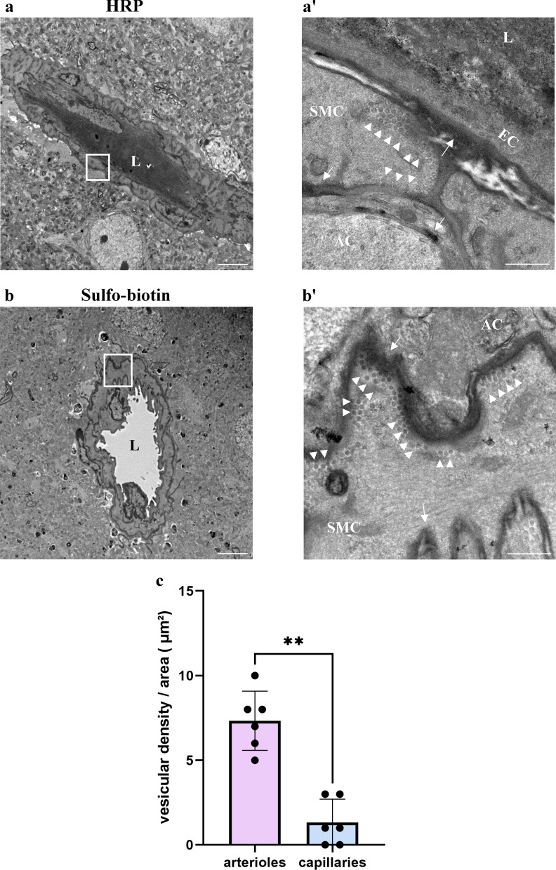
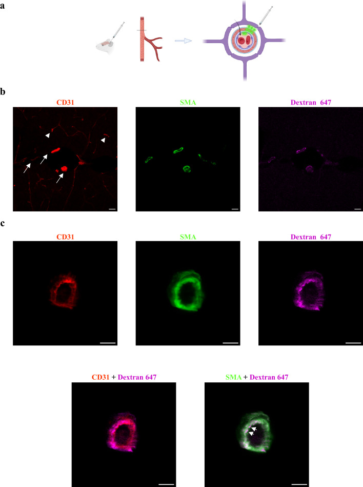
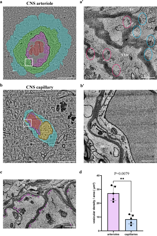
CNS vasculature differs from vascular networks of peripheral organs by its ability to tightly control selective material exchange across capillary barriers. Capillary permeability is mostly defined by unique cellular components of the endothelium. While capillaries are extensively investigated, the barrier properties of larger vessels are understudied. Here, we investigate barrier properties of CNS arterial walls. Using tracer challenges and various imaging modalities, we discovered that at the mouse cortex, the arterial barrier does not reside at the classical level of the endothelium. The arterial wall's unique permeability acts bi-directionally; CSF substances travel along the glymphatic path and can penetrate from the peri-vascular space through arteriolar walls towards the lumen. We found that caveolae vesicles in arteriole endothelial are functional transcytosis machinery components, and that a similar mechanism is evident in the human brain. Our discoveries highlight vascular heterogeneity investigations as a potent approach to uncover new barrier mechanisms.

Keywords: Arterial berrier; BBB; Super-resolution; Transcytosis; dSTORM.


Conflict of interest statement

The authors declare no competing interests.

Figures

Fig. 1
Fig. 1
Variable degree of permeability to different tracers across the CNS arteriole-wall uncovers atypical cellular barrier properties. Confocal-microscopy of arteriole-wall permeability, with immunostaining for NVU components: endothelium (anti-CD31), smooth-muscle (anti-SMA), astrocyte end-feet (anti-AQP4), and basement membranes (anti-pan Laminin) of wild-type adult mouse cortical sections. a Schematic illustration of the experimental design: tracers of different size and molecular compositions are introduced into the blood stream and circulated for 10 min. Arteriole cross sections are used to locate fluorescent tracer signals (green circles) in the vessel wall from luminal side (blood) towards the tissue (arrow showing the path crossing the endothelial cell layer-purple, smooth-muscle layer-red each surrounded by basement membranes and all surrounded by the astrocyte end-feet) Created with BioRender.com. b Tracer challenges with Alexa-647 conjugated albumin for 10 min (70 kDa) demonstrating that albumin is mostly co-localized with the endothelium (arrow). c Challenges with Alexa-647 conjugated dextran for 10 min (10 kDa) demonstrates tracer signals co-localized with the endothelium and reaching the smooth-muscle layer (upper panel, arrow), and beyond reaching the astrocyte end-feet (lower panel, arrow). d Challenges with perfused sulfo-biotin (443 Da), stained with Alexa-647 conjugated streptavidin demonstrates tracer signals passed the smooth-muscle layer (upper panel, arrow), but confined by the basement membranes (lower panel, arrow). Images are representative of n = 27 arterioles profiles of n = 9 mice (3 for each tracer), Scale Bars 10 µm
Fig. 2
Fig. 2
dSTORM imaging demonstrates super-resolution tracer permeability and validates the unique barrier properties of the CNS arteriole wall. Precise nano-scale localization of the sulfo-biotin tracer (443 Da), stained with Alexa-647 conjugated streptavidin. a Relatively low resolution TIRF mode imaging (Total Internal Reflection Fluorescence, similar to epi-fluorescence with TIRF illumination, presenting all the collected signals with no super-resolution analysis) of endothelium (anti-CD31), smooth-muscle (anti-SMA) in wild-type adult mouse brain sections, does not allow precise localization of tracer along the vascular wall due to diffraction limitation. Scale bar 10 µm. b dSTORM images of the same arteriole (as in a) shows that tracer signals are found passed both the endothelium and the smooth-muscle markers. Scale bar 10 µm. b’-b’’, Inset magnifications showing distances between the tracer and the cell markers (CD31 (b’, SMA signal omitted) and of SMA (b’’), scale bars b’ 2 µm, b’’ 1 µm). c Quantification of tracer signals beyond the smooth muscle marker (sulfo-biotin tracer images appear here, dextran and albumin tracers images appear in Additional file 1: Fig. S1). There are no significant differences in permeability of these three tracers of different size and molecular compositions (Kruskal–Wallis H test). (L) marks the vessel lumen. Dashed arrows marks tracer direction from the lumen towards the parenchyma. Images are representative of n = 18 arteriole profiles of n = 4 mice
Fig. 3
Fig. 3
Caveolae vesicles in arteriole endothelial and smooth muscle cells are functional transcytosis machinery components. HRP and sulfo-biotin tracers, imaged by TEM, demonstrate cargo trafficking in CNS arteriole cells. a Representative TEM image of a cortical arteriole (wild-type adult mice) following 30 min HRP tracer challenge. HRP signal is found in vessel lumen (L) as well as in basement membranes; in between the endothelial layer and smooth muscle layer (arrows), and between the smooth-muscle layer and astrocyte end-feet (arrows). Scale bar 5 µm. Inset (a’) ample caveolae vesicles, some of which showing HRP signals at luminal and abluminal membranes of a smooth muscle cell (arrowheads). Scale bar 500 nm. b Representative TEM image of perfused sulfo-biotin tracer challenge following staining with HRP conjugated streptavidin. Scale bar 5 µm. Inset (b’) high magnification image showing abundant tracer-field vesicles, adjacent to the abluminal membrane of a smooth-muscle cell (arrowheads). Tracer signal is found also in basement membranes (arrows). SMC Smooth-muscle cell, EC Endothelial cell, AC Astrocyte, L lumen. Scale bar 500 nm (n = 6 mice, 12 arterioles). c Vesicular density in mouse arterial endothelial cells compared to capillary endothelial cells. Mean vesicular density in cECs and aECs. Data are mean ± s.e.m. **p < 0.05 (Two tailed Mann–Whitney U- test)
Fig. 4
Fig. 4
Tracer introduced into the CSF penetrates into the perivascular space of arteriolar wall. For testing the potential of clearance mechanisms, we adopted the methodology used to study the ‘Glymphatic’ path, and injected Dextran (10 kDa) into the cisterna magna. a Schematic illustration of the experimental design: very small volumes of tracers are introduced into the cisterna magna and circulate in the CSF for 30 min. Cortical cross sections are used to locate fluorescent tracer signals (green circles). b Low magnification confocal microscopy of cortical sections demonstrates expected tracer signals around arteries/arterioles (arrows, CD31 + SMA double positive vessels), but not around capillaries (arrowheads, CD31 single positive vessels). Scale bar 5 µm c, Confocal microscopy imaging of an arteriole cross section demonstrates approximate co-localization of the tracer with both endothelium and smooth-muscle signals (arrows). Scale bar 10 µm (n = 3 mice, 12 arterioles)
Fig. 5
Fig. 5
Arterial wall permeability is bi-directional. Tracer introduced into the CSF travel along the Glymphatic path and can penetrate from the perivascular space across arteriolar walls towards the luminal direction. a, b 30 min CSF tracer challenges with Alexa-647 conjugated dextran (10 kDa, cisterna magna injections) and immunostaining for SMA and CD31 of wild-type adult brain sections. (L) marks the vessel lumen. Dashed arrows marks tracer direction towards the lumen. Scale bars 10 µm a Precise nano-scale localization with dSTORM imaging shows tracer signals located along the CSF-blood trajectory. Inset (a’ scale bars 2 µm) magnifications demonstrating tracer signals between smooth-muscle and endothelium markers (arrows), and at the endothelium luminal side (arrowhead). b Based on tracer signal distribution, especially at high magnification (inset b’ scale bars 100 nm), we identify tracer clusters that fit the dimensions of transcytosis vesicles. These vesicle-like structures are located in smooth muscle cells (arrowheads, in 1 and 2). Elongated distribution might represent tracer filled basement membrane between the smooth muscle and the endothelial cell (arrow, in 2). Vesicle-shaped structures that are connected to the basement membrane resemble the ultrastructure of flask shape membrane pits (astrix); n = 3 mice, 12 arterioles
Fig. 6
Fig. 6
Abundant vesicles in arteriole cells of human CNS might indicate transcytotic activity. Caveolae vesicles in arteriole endothelial and smooth muscle cells are evident in 3D EM reconstruction of human cortical tissue [14]. a, b Transmission electron microscopy images of a CNS arteriole and capillary. Pseudo-colors highlight cell types: smooth-muscle (green), endothelium (purple), pericyte (yellow), astrocyte end-foot (blue), red blood cell (red), Scale bar 10 µm. a’, b’, Magnification of the boxed area in the left panel. a’ Abundant vesicles in aEC’s (blue ovals) and aSMCs (red ovals). c, Example of multiple vesicles (pseudo labeled in magenta) in aSMCs. d Quantification showing significant higher vesicular density in aECs than in cECs (n = 5 arterioles and 5 capillaries). SMC Smooth-muscle cell, EC Endothelial cell, AC Astrocyte, P Pericyte, L lumen. Scale bar 1 µm. Data are mean ± s.e.m. ** p < 0.05 (two tailed Mann–Whitney U- test)

References

    1. Reese TS, Karnovsky MJ. Fine structural localization of a blood-brain barrier to exogenous peroxidase. J Cell Biol. 1967;34:207–217. doi: 10.1083/jcb.34.1.207. - DOI - PMC - PubMed
    1. Langen UH, Ayloo S, Gu C. Development and cell biology of the blood-brain barrier. Annu Rev Cell Dev Biol. 2019;35:591–613. doi: 10.1146/annurev-cellbio-100617-062608. - DOI - PMC - PubMed
    1. Ben-Zvi A, et al. Mfsd2a is critical for the formation and function of the blood-brain barrier. Nature. 2014;509:507–511. doi: 10.1038/nature13324. - DOI - PMC - PubMed
    1. Licht T, et al. Hippocampal neural stem cells facilitate access from circulation via apical cytoplasmic processes. Elife. 2020 doi: 10.7554/eLife.52134. - DOI - PMC - PubMed
    1. Pulido RS, et al. Neuronal activity regulates blood-brain barrier efflux transport through endothelial circadian genes. Neuron. 2020;108:937–952.e937. doi: 10.1016/j.neuron.2020.09.002. - DOI - PMC - PubMed

LinkOut - more resources