

An Inverse Method to Determine Mechanical Parameters of Porcine Vitreous Bodies Based on the Indentation Test
- PMID: 37370577
- PMCID: PMC10295402
- DOI: 10.3390/bioengineering10060646
An Inverse Method to Determine Mechanical Parameters of Porcine Vitreous Bodies Based on the Indentation Test
Abstract
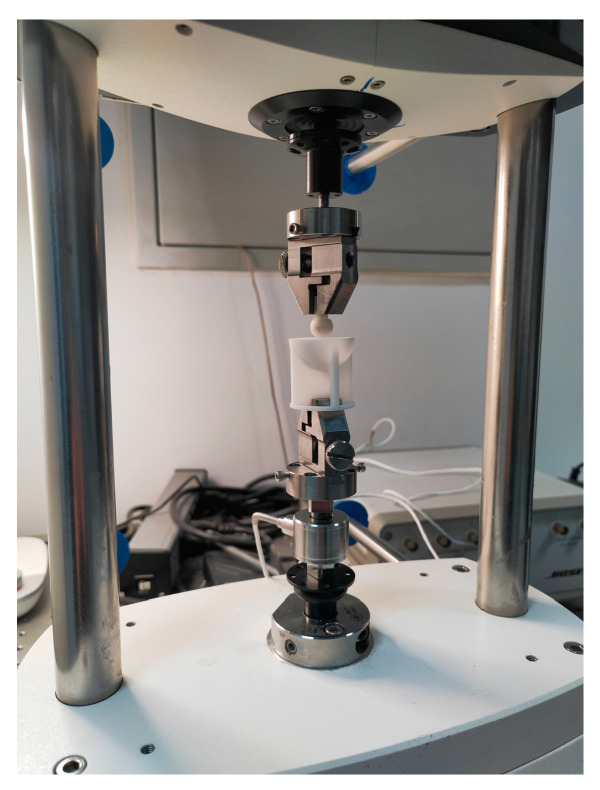
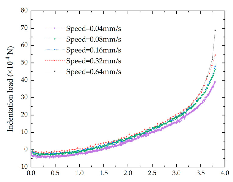
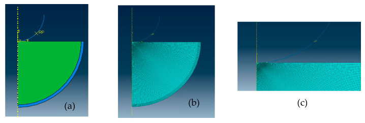
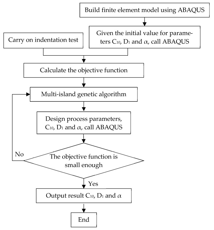
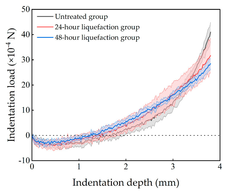
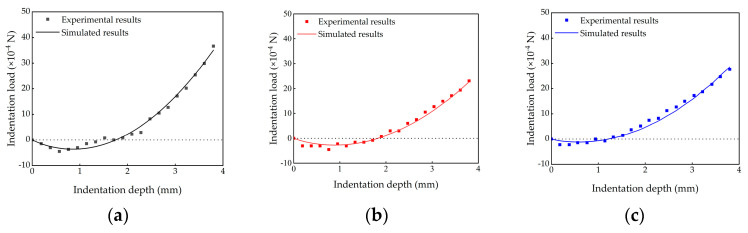
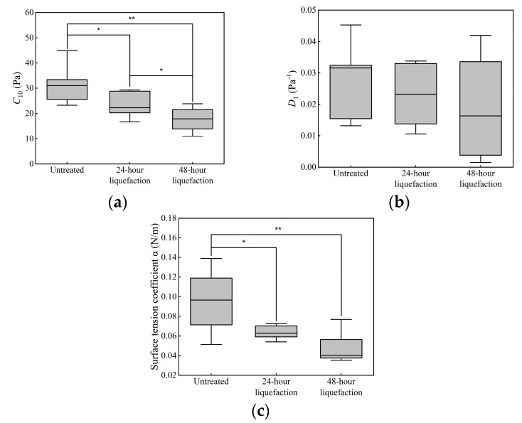
The vitreous body keeps the lens and retina in place and protects these tissues from physical insults. Existing studies have reported that the mechanical properties of vitreous body varied after liquefaction, suggesting mechanical properties could be effective parameters to identify vitreous liquefaction process. Thus, in this work, we aimed to propose a method to determine the mechanical properties of vitreous bodies. Fresh porcine eyes were divided into three groups, including the untreated group, the 24 h liquefaction group and the 48 h liquefaction group, which was injected collagenase and then kept for 24 h or 48 h. The indentation tests were carried out on the vitreous body in its natural location while the posterior segment of the eye was fixed in the container. A finite element model of a specimen undertaking indentation was constructed to simulate the indentation test with surface tension of vitreous body considered. Using the inverse method, the mechanical parameters of the vitreous body and the surface tension coefficient were determined. For the same parameter, values were highest in the untreated group, followed by the 24 h liquefaction group and the lowest in the 48 h liquefaction group. For C10 in the neo-Hookean model, the significant differences were found between the untreated group and liquefaction groups. This work quantified vitreous body mechanical properties successfully using inverse method, which provides a new method for identifying vitreous liquefactions related studies.
Keywords: indentation test; inverse method; liquefaction; mechanical properties; vitreous body.
Conflict of interest statement
The authors declare no conflict of interest.
Figures
References
Grants and funding
LinkOut - more resources
Full Text Sources
