Review
. 2023 Jun 3;10(6):682.
doi: 10.3390/bioengineering10060682.

Polysaccharides and Structural Proteins as Components in Three-Dimensional Scaffolds for Breast Cancer Tissue Models: A Review

Affiliations
Review

Polysaccharides and Structural Proteins as Components in Three-Dimensional Scaffolds for Breast Cancer Tissue Models: A Review

Eva Pasquier et al. Bioengineering (Basel). .

Abstract

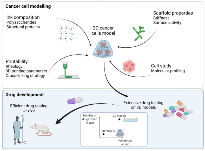
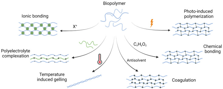
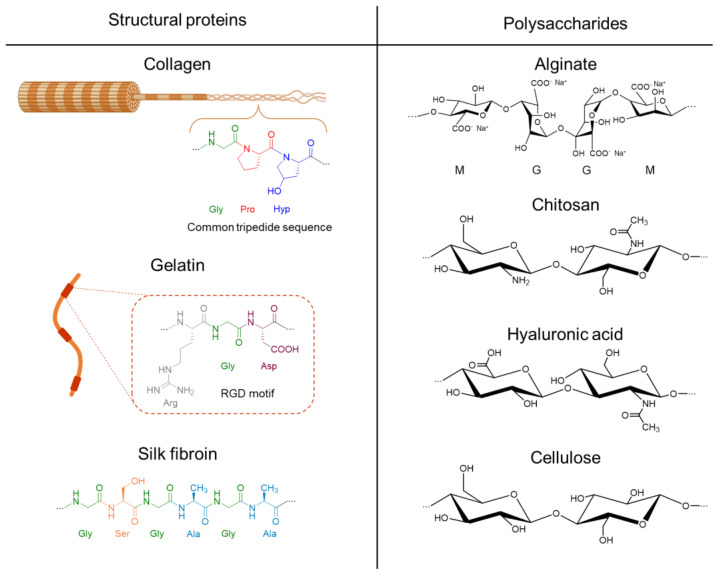
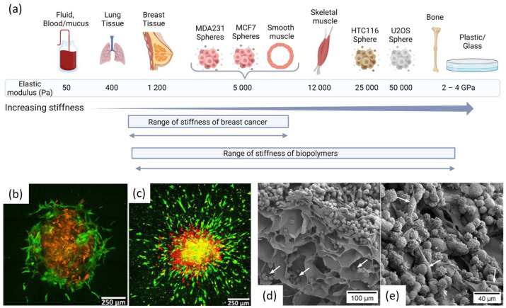
Breast cancer is the most common cancer among women, and even though treatments are available, efficiency varies with the patients. In vitro 2D models are commonly used to develop new treatments. However, 2D models overestimate drug efficiency, which increases the failure rate in later phase III clinical trials. New model systems that allow extensive and efficient drug screening are thus required. Three-dimensional printed hydrogels containing active components for cancer cell growth are interesting candidates for the preparation of next generation cancer cell models. Macromolecules, obtained from marine- and land-based resources, can form biopolymers (polysaccharides such as alginate, chitosan, hyaluronic acid, and cellulose) and bioactive components (structural proteins such as collagen, gelatin, and silk fibroin) in hydrogels with adequate physical properties in terms of porosity, rheology, and mechanical strength. Hence, in this study attention is given to biofabrication methods and to the modification with biological macromolecules to become bioactive and, thus, optimize 3D printed structures that better mimic the cancer cell microenvironment. Ink formulations combining polysaccharides for tuning the mechanical properties and bioactive polymers for controlling cell adhesion is key to optimizing the growth of the cancer cells.

Keywords: 3D bioprinting; biopolymers; breast cancer models; cells microenvironment.


Conflict of interest statement

A.S. (Anders Ståhlberg) declares stock ownership and is a board member in Tulebovaasta, Iscaff Pharma and SiMSen Diagnostics AB.

Figures

Figure 6
Figure 6
Schematic overview of gene expression profiling using 3D models. To analyze the gene expression profile of all cells, RNA is extracted from either the entire 3D model or the harvested cells followed by reverse transcription and either qPCR or sequencing. Direct lyzed cells from the scaffold can also be transferred directly to the reverse transcription step without any extraction [155]. For single cell gene expression profiling, individual cells need to be collected from the 3D model followed by reverse transcription and either qPCR or sequencing. Created with BioRender.com.
Figure 1
Figure 1
Steps to make a 3D cancer cell model by combining different biopolymers and cells to make a bioink for 3D printing. After optimization of the cancer cells microenvironment screening of high number of drugs is possible before selecting the most efficient drugs for in vivo testing, hence reducing the number and improving the success rate of drugs tested in vivo. Created with BioRender.com.
Figure 2
Figure 2
3D printing of complex structures for in vitro tumor modeling. (a) 3D bioprinting of a heterogeneous tumor model comprised of both MD Anderson-Metastatic Breast-231 Cells (MDA-MB-231) breast cancer cells and Institute for Medical Research-90 (IMR-90) fibroblasts to study cells migration and interactions. Model and photograph of the bioprinted sample. Reproduced with permission from [32] (b) 3D printing of a vascularized tissue model for studying breast cancer metastasis to the bone. Schematic of printed model and images of the bone and tumor regions. Images of 3D printed sample with top and side view. Reproduced with permission from [35].
Figure 3
Figure 3
Different types of cross-linking commonly used to stabilize 3D printed biopolymer structures. Created with BioRender.com.
Figure 4
Figure 4
Structure of relevant proteins and polysaccharides that can form 3D scaffolds that support cancer cell growth: collagen, gelatin, silk fibroin, and alginate on the sodium form, chitosan, hyaluronic acid, and cellulose.
Figure 5
Figure 5
(a) Elastic modulus of various tissues, including normal breast tissue and breast cancer tissue. Data extracted from [22,96] and figure created with BioRender.com b. and c. Tumor spheroid invasion in 3D gel containing only collagen (b) and alginate and collagen (c) A spheroid transferred into a 3D gel at day 0 and cultured for 6 days in the gel shows minimal invasion only of human mammary fibroblasts cells in collagen and extensive invasion in alginate-collagen gel. Red color illustrates MDA-MB-231 cells expressing mKate fluorescent protein and green color illustrates human mammary fibroblasts cells expressing green fluorescent protein. Reproduced with permission from [95]. (d,e) MDA-231 cells growing on nanocellulose scaffolds. d. Cross-sectional image of a 3D scaffold. Note the cells reaching the pores of the scaffold (white arrows). (e) A heterogeneous population of MDA-231 cells (round and elongated cells indicated by white arrows), growing on the surface the scaffold.

References

    1. Sung H., Ferlay J., Siegel R.L., Laversanne M., Soerjomataram I., Jemal A., Bray F. Global Cancer Statistics 2020: GLOBOCAN Estimates of Incidence and Mortality Worldwide for 36 Cancers in 185 Countries. CA A Cancer J. Clin. 2021;71:209–249. doi: 10.3322/caac.21660. - DOI - PubMed
    1. Bray F., Ferlay J., Soerjomataram I., Siegel R.L., Torre L.A., Jemal A. Global cancer statistics 2018: GLOBOCAN estimates of incidence and mortality worldwide for 36 cancers in 185 countries. CA A Cancer J. Clin. 2018;68:394–424. doi: 10.3322/caac.21492. - DOI - PubMed
    1. Lloyd I. Pharma R&D Annual Review 2019. Pharma Intelligence; London, UK: 2019.
    1. Perrin S. Preclinical research: Make mouse studies work. Nature. 2014;507:423–425. doi: 10.1038/507423a. - DOI - PubMed
    1. Hutchinson L., Kirk R. High drug attrition rates—Where are we going wrong? Nat. Rev. Clin. Oncol. 2011;8:189–190. doi: 10.1038/nrclinonc.2011.34. - DOI - PubMed

LinkOut - more resources