

Spatial Transcriptomics Identifies Expression Signatures Specific to Lacrimal Gland Adenoid Cystic Carcinoma Cells
- PMID: 37370820
- PMCID: PMC10296284
- DOI: 10.3390/cancers15123211
Spatial Transcriptomics Identifies Expression Signatures Specific to Lacrimal Gland Adenoid Cystic Carcinoma Cells
Abstract
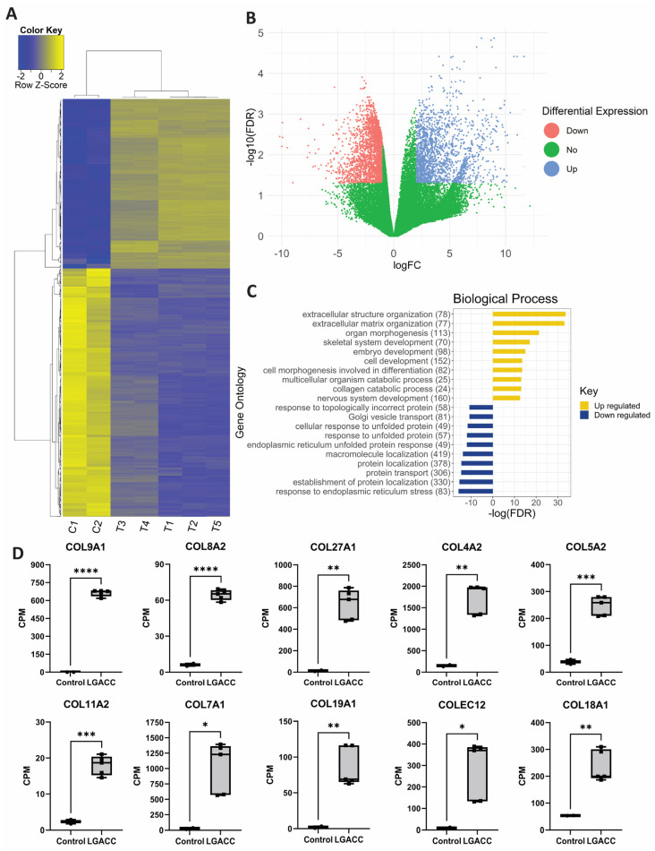
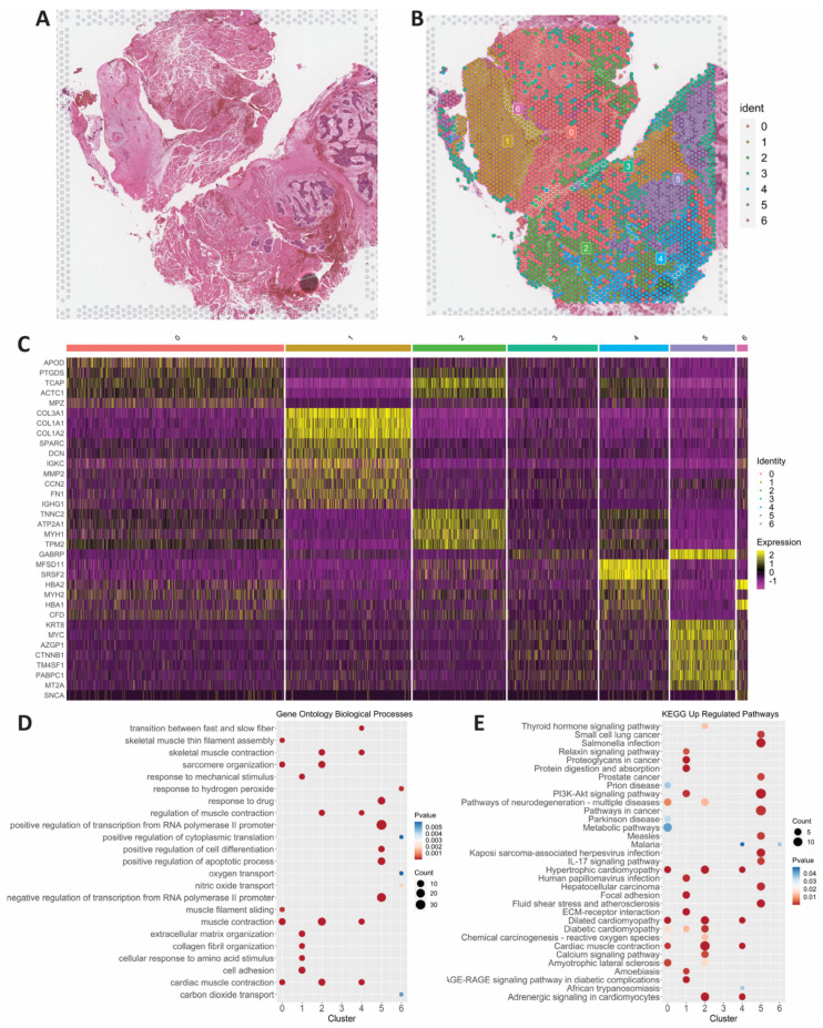
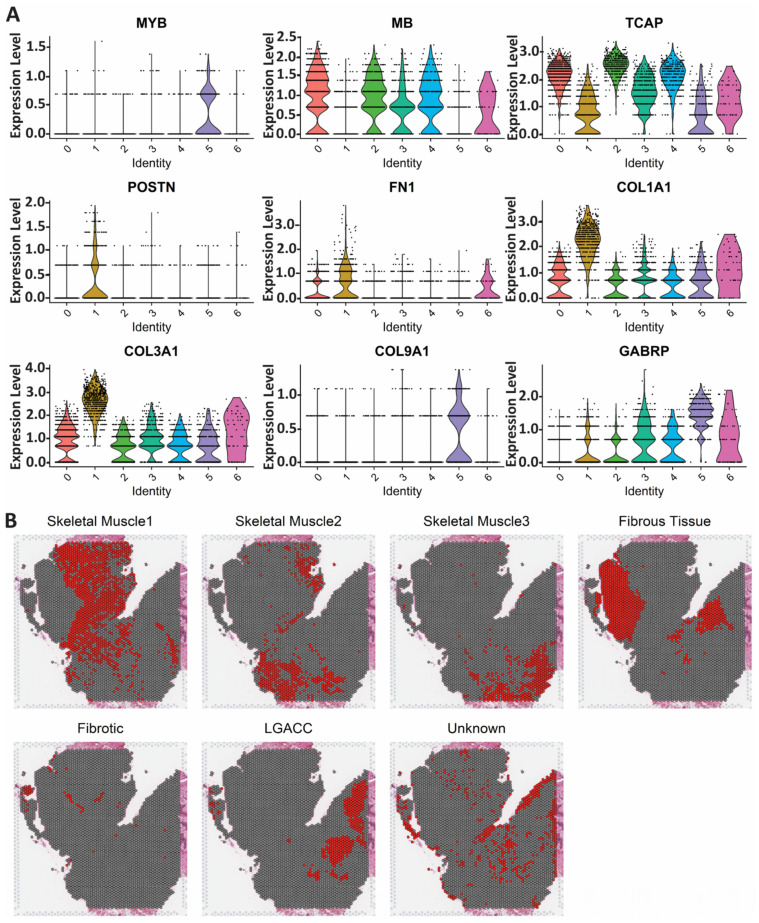
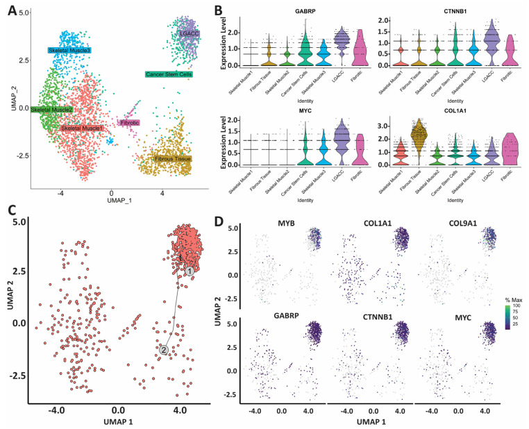
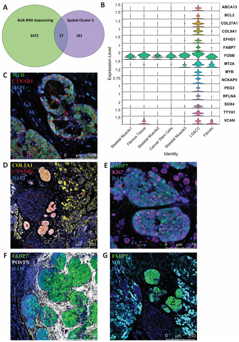
Although primary tumors of the lacrimal gland are rare, adenoid cystic carcinoma (ACC) is the most common and lethal epithelial lacrimal gland malignancy. Traditional management of lacrimal gland adenoid cystic carcinoma (LGACC) involves the removal of the eye and surrounding socket contents, followed by chemoradiation. Even with this radical treatment, the 10-year survival rate for LGACC is 20% given the propensity for recurrence and metastasis. Due to the rarity of LGACC, its pathobiology is not well-understood, leading to difficulties in diagnosis, treatment, and effective management. Here, we integrate bulk RNA sequencing (RNA-seq) and spatial transcriptomics to identify a specific LGACC gene signature that can inform novel targeted therapies. Of the 3499 differentially expressed genes identified by bulk RNA-seq, the results of our spatial transcriptomic analysis reveal 15 upregulated and 12 downregulated genes that specifically arise from LGACC cells, whereas fibroblasts, reactive fibrotic tissue, and nervous and skeletal muscle account for the remaining bulk RNA-seq signature. In light of the analysis, we identified a transitional state cell or stem cell cluster. The results of the pathway analysis identified the upregulation of PI3K-Akt signaling, IL-17 signaling, and multiple other cancer pathways. This study provides insights into the molecular and cellular landscape of LGACC, which can inform new, targeted therapies to improve patient outcomes.
Keywords: lacrimal gland adenoid cystic carcinoma; rare cancer; spatial transcriptomics; transcriptomic signature.
Conflict of interest statement
The authors declare no conflict of interest. The funders had no role in the design of the study; in the collection, analyses, or interpretation of data; in the writing of the manuscript; or in the decision to publish the results.
Figures
Similar articles
-
Mass spectrometry database of lacrimal gland adenoid cystic carcinoma and normal lacrimal gland tissue identifies extracellular matrix remodeling in these tumors.Data Brief. 2023 Jun 19;49:109330. doi: 10.1016/j.dib.2023.109330. eCollection 2023 Aug. Data Brief. 2023. PMID: 37409171 Free PMC article.
-
Treatment strategies and prognostic insights for lacrimal gland adenoid cystic carcinoma: a review.Discov Oncol. 2025 May 22;16(1):858. doi: 10.1007/s12672-025-02468-5. Discov Oncol. 2025. PMID: 40402353 Free PMC article. Review.
-
Whole Exome Sequencing of Lacrimal Gland Adenoid Cystic Carcinoma.Invest Ophthalmol Vis Sci. 2017 May 1;58(6):BIO240-BIO246. doi: 10.1167/iovs.16-21097. Invest Ophthalmol Vis Sci. 2017. PMID: 28820917 Free PMC article.
-
Neoadjuvant Intra-arterial Cytoreductive Chemotherapy Improves Outcomes in Lacrimal Gland Adenoid Cystic Carcinoma.Oncologist. 2024 Mar 4;29(3):263-269. doi: 10.1093/oncolo/oyad346. Oncologist. 2024. PMID: 38227581 Free PMC article.
-
A Review of the Molecular Landscape of Adenoid Cystic Carcinoma of the Lacrimal Gland.Int J Mol Sci. 2023 Sep 6;24(18):13755. doi: 10.3390/ijms241813755. Int J Mol Sci. 2023. PMID: 37762061 Free PMC article. Review.
Cited by
-
Circulating Adenoid Cystic Carcinoma associated MYB transcripts enable rapid and sensitive detection of metastatic disease in blood liquid biopsies.J Liq Biopsy. 2024 Dec;6:100276. doi: 10.1016/j.jlb.2024.100276. J Liq Biopsy. 2024. PMID: 39801676 Free PMC article.
-
Prognostic factors for lacrimal gland adenoid cystic carcinoma: a retrospective study in Chinese patients.Int J Ophthalmol. 2024 Aug 18;17(8):1423-1430. doi: 10.18240/ijo.2024.08.06. eCollection 2024. Int J Ophthalmol. 2024. PMID: 39156780 Free PMC article.
-
Circulating Adenoid Cystic Carcinoma associated MYB transcripts enable rapid and sensitive detection of metastatic disease in blood liquid biopsies.medRxiv [Preprint]. 2024 Oct 17:2024.10.15.24315549. doi: 10.1101/2024.10.15.24315549. medRxiv. 2024. Update in: J Liq Biopsy. 2024 Dec;6:100276. doi: 10.1016/j.jlb.2024.100276. PMID: 39484279 Free PMC article. Updated. Preprint.
-
Treatment of lacrimal gland adenoid cystic carcinoma: a systematic review and Meta-analysis.Int J Ophthalmol. 2024 Jan 18;17(1):164-172. doi: 10.18240/ijo.2024.01.22. eCollection 2024. Int J Ophthalmol. 2024. PMID: 38239951 Free PMC article.
-
Intraoperative use of electrical impedance spectroscopy for adenoid cystic carcinoma of the lacrimal gland: A case report.Clin Case Rep. 2023 Oct 9;11(10):e7995. doi: 10.1002/ccr3.7995. eCollection 2023 Oct. Clin Case Rep. 2023. PMID: 37822486 Free PMC article.
References
Grants and funding
LinkOut - more resources
Full Text Sources
Molecular Biology Databases
