. 2023 Jun 18;15(12):3234.
doi: 10.3390/cancers15123234.

Effect of Mitotane on Male Gonadal Function

Affiliations

Effect of Mitotane on Male Gonadal Function

Federica Innocenti et al. Cancers (Basel). .

Abstract

Background: Clinical evidence has shown frequent hypogonadism following mitotane (MTT) treatment in male patients with adrenocortical carcinoma. This study aimed to evaluate the impact of MTT on male gonadal function.

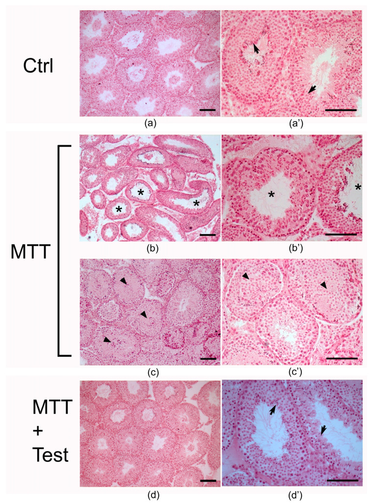
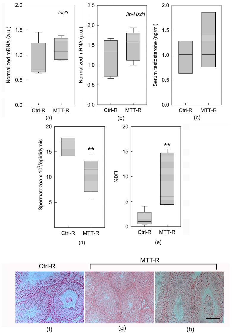
Methods: Morphological analysis of testes and testosterone assays were performed on adult CD1 MTT-treated and untreated mice. The expression of key genes involved in interstitial and tubular compartments was studied by real-time PCR. Moreover, quantitative and qualitative analysis of spermatozoa was performed.

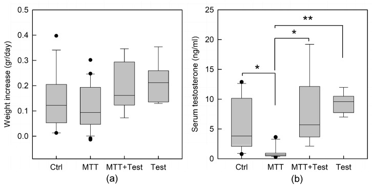
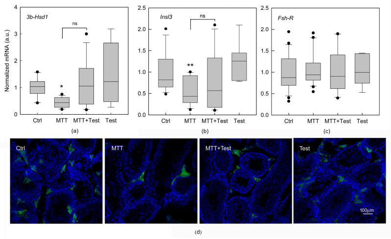
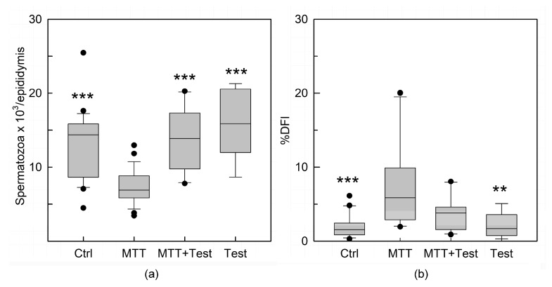
Results: Several degrees of damage to the testes and a significant testosterone reduction in MTT-treated mice were observed. A significant decline in 3βHsd1 and Insl3 mRNA expression in the interstitial compartment confirmed an impairment of androgen production. Fsh-R mRNA expression was unaffected by MTT, proving that Sertoli cells are not the drug's primary target. Sperm concentrations were significantly lower in MTT-treated animals. Moreover, the drug caused a significant increase in the percentage of spermatozoa with abnormal chromatin structures.

Conclusion: MTT negatively affects the male reproductive system, including changes in the morphology of testicular tissue and reductions in sperm concentration and quality.

Keywords: adrenocortical carcinoma; male gonadal function; mitotane; testis; testosterone.


Conflict of interest statement

The authors declare no conflict of interest.

Figures

Figure 1
Figure 1
Effect of mitotane on: (a) daily body weight increase; and (b) serum testosterone dosage in control (Ctrl), mitotane (MTT), MTT-testosterone cotreated (MTT + Test), and testosterone (Test) treated animals. The values are expressed as the mean ± s.e.m. with a total animal number of C = 10; MTT = 11; M + T = 6; Test = 6. Statistical analysis was performed by one-way ANOVA, followed by the Tukey–Kramer test. * p < 0.05; ** p < 0.01.
Figure 2
Figure 2
Photomicrographs of sections of testes from (a,a’, Ctrl) control, (bc’, MTT) MTT-treated, and (d,d’, MTT + Test) MTT and testosterone-treated mice. Arrows: spermatozoa; asterisks: tubules with enlarged lumen; arrowheads: tubules with absence of lumen. Scale bar = 100 μm.
Figure 3
Figure 3
Effects of mitotane on gene expression in isolated seminiferous tubules and Leydig cells removed from untreated mice (Ctrl) and mice treated with mitotane (MTT), MTT and testosterone (MTT+Test) together, and testosterone (Test). Expression of 3β-Hsd1 (a) and Insl3 (b) in LCs, detected by real-time PCR in LCs within interstitial cells. Expression of Fsh-R (c) mRNA in mouse SCs within the seminiferous tubules detected by real-time PCR. Each sample was normalized to its β-actin content. Results are expressed as arbitrary units (a.u.) and are represented as the mean ± s.e.m. of three independent experiments with total animal numbers of Ctrl = 15, MTT = 18 and MTT + Test = 12, and testosterone = 7. Statistical analysis was performed using ANOVA followed by the Tukey–Kramer test; * p < 0.05 and ** p < 0.01, ns: no Significance. Immunofluorescence analysis (d). Representative images of immunofluorescent 3β-HSD staining in sections of testes from mice treated as described above. Scale bar = 100 μm.
Figure 4
Figure 4
Mean of the number of spermatozoa in mice injected with DMSO (Ctrl), MTT, MTT and testosterone together (MTT + Test) and testosterone (Test). Results are expressed as number of spermatozoa per epididymis (a). Quantification of sperm with abnormal chromatin structures calculated by the evaluation of the parameter DNA fragmentation index (DFI). A high %DFI correspond to a large percentage of damaged germ cells. Results are expressed as arbitrary units (a.u.) (b). Values are represented as the mean ± s.e.m. of three independent experiments with total animal numbers of Ctrl = 12, MTT = 12, MTT + Test = 7 and Test = 4. Statistical analysis was performed using ANOVA followed by the Tukey–Kramer test. ** p < 0.01, *** p < 0.001 vs. MTT.
Figure 5
Figure 5
Effect of MTT withdrawal on the expression of Insl3 (a) and 3β-Hsd1 (b) in LCs removed from untreated (Ctrl) and mitotane (MTT)-treated mice at 30 days after MTT removal, detected by real-time PCR in LCs within interstitial cells. Each sample was normalized to its β-actin content. Results are expressed as arbitrary units (a.u.) and are represented as the mean ± s.e.m. of three independent experiments with a total animal number of Ctrl = 6 and MTT = 6. Serum testosterone concentration (c). Mean of the number of spermatozoa (d). Quantification of sperm with abnormal chromatin structures calculated by the evaluation of the parameter DNA fragmentation index (DFI) (e). A high %DFI corresponds to a large percentage of damaged germ cells. Statistical analysis was performed using Student’s t test. ** p < 0.01 vs. respective Ctrl-R. Testis morphology after MTT withdrawal (fh). Scale bar = 100 mm.

References

    1. Fassnacht M., Assie G., Baudin E., Eisenhofer G., de la Fouchardiere C., Haak H.R., de Krijger R., Porpiglia F., Terzolo M., Berruti A. Adrenocortical carcinomas and malignant phaeochromocytomas: ESMO-EURACAN Clinical Practice Guidelines for diagnosis, treatment and follow-up. Ann. Oncol. 2020;31:1476–1490. doi: 10.1016/j.annonc.2020.08.2099. - DOI - PubMed
    1. Barbot M., Zilio M., Scaroni C. Cushing’s syndrome: Overview of clinical presentation, diagnostic tools and complications. Best Pract. Res. Clin. Endocrinol. Metab. 2020;34:101380. doi: 10.1016/j.beem.2020.101380. - DOI - PubMed
    1. Stigliano A., Chiodini I., Giordano R., Faggiano A., Canu L., Della Casa S., Loli P., Luconi M., Mantero F., Terzolo M. Management of adrenocortical carcinoma: A consensus statement of the Italian Society of Endocrinology (SIE) J. Endocrinol. Investig. 2016;39:103–121. doi: 10.1007/s40618-015-0349-9. - DOI - PubMed
    1. Baudry C., Coste J., Bou Khalil R., Silvera S., Guignat L., Guibourdenche J., Abbas H., Legmann P., Bertagna X., Bertherat J. Efficiency and tolerance of mitotane in Cushing’s disease in 76 patients from a single center. Eur. J. Endocrinol. 2012;167:473–481. doi: 10.1530/EJE-12-0358. - DOI - PubMed
    1. Innocenti F., Cerquetti L., Pezzilli S., Bucci B., Toscano V., Canipari R., Stigliano A. Effect of mitotane on mouse ovarian follicle development and fertility. J. Endocrinol. 2017;234:29–39. doi: 10.1530/JOE-17-0203. - DOI - PubMed

LinkOut - more resources