Review
. 2023 Jun 4;10(6):1012.
doi: 10.3390/children10061012.

Hybrid Palliation for Hypoplastic Left Heart Syndrome: Role of Echocardiography

Affiliations
Review

Hybrid Palliation for Hypoplastic Left Heart Syndrome: Role of Echocardiography

Lilia Oreto et al. Children (Basel). .

Abstract

Hypoplastic left heart syndrome is a spectrum of complex congenital cardiac defects. Although in borderline cases, biventricular repair is a viable option, in the majority of cases, univentricular palliation is the treatment of choice. Hybrid palliation can be a valid alternative to classic Norwood operation in the neonatal period, especially in selected cases such as high-risk patients or borderline left ventricles. Echocardiography is the main diagnostic modality in this pediatric population, from the fetal diagnosis to the subsequent surgical steps of palliative treatment. Hybrid palliation is performed after birth and is characterized by surgical banding of the pulmonary arteries along with transcatheter stenting of the ductus arteriosus. There are some peculiar aspects of cardiac imaging that characterize this type of palliation, and that should be considered in the different phases before and after the procedure. We aimed to review the current literature about the role of echocardiography in the management of patients with hypoplastic left heart undergoing hybrid palliation.

Keywords: echocardiography; hybrid palliation; hypoplastic left heart syndrome.


Conflict of interest statement

The authors declare no conflict of interest.

Figures

Figure 1
Figure 1
Fetal echocardiography. Reverse flow in the aortic arch is shown from a long-axis view of the aortic arch and ductal arch (panel (A)) and from a three-vessel view (panel (B)). Panel (C) shows severe hypoplasia of all the segments of the aortic arch with retrograde flow. Four-chamber view in panel (D) shows mitral valve stenosis with hypoplastic left ventricle, which does not form cardiac apex. In the four-chamber view in panel (E) mitral valve is atretic, and the left ventricle is a virtual chamber. Tricuspid valve with thickened leaflets and severe regurgitation is shown in Panel (F). In panel (G), the mitral valve is highly dysplastic, with a very small opening in diastole. Panel (H) shows a left-to-right shunt through the foramen ovale with inverted movement of the membrane. A vertical vein draining into the left atrium is shown in Panel (I).
Figure 2
Figure 2
Dilated pulmonary trunk with aortic valve atresia and wide ductus arteriosus providing systemic cardiac output with right-to-left shunt in systole (panel (A)) and left-to-right shunt due to lower pulmonary compared to systemic resistances (panel (B)). Continous-wave Doppler through the ductus allows measurement of the amount of flow in each direction by tracing the Doppler curves (Panel (C)). Panel (D) shows a diminutive takeoff of the aortic arch from the ductus, generating increased velocity of the reversed aortic flow measured by Pulsed-wave Doppler (Panel (E)). Ductus arteriosus can be very large compared to pulmonary arteries, as shown in panel (F).
Figure 3
Figure 3
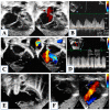
Atrial septal defect with malaligned septum primum and no restrictive flow (Panel (A)). Continous wave Doppler showing a low mean gradient of 2 mmHg through the atrial septum (Panel (B)). Unrestrictive ostium secundum atrial septal defect (Panel (C)). Critically small atrial septal communication with aliasing by color-Doppler (Panel (D)) and high mean gradient of 12 mmHg (Panel (E)). Successful stenting of the restrictive atrial defect (Panel (F)).
Figure 4
Figure 4
Evaluation of right ventricular function. Fractional area change of the right ventricle is obtained by tracing the endocardial border in diastole (panel (A)) and in systole (panel (B)). Tissue Doppler imaging of the lateral tricuspid annulus allows the measurement of the annular systolic velocity (panel (C)). Tricuspid annular plane systolic excursion is obtained by M-Mode (Panel (D)). Mild tricuspid regurgitation (panel (E)). Continous-wave Doppler of the tricuspid regurgitation jet allows measurement of the systolic to diastolic duration ratio (Panel (F)).
Figure 5
Figure 5
Severe aortic valve stenosis is shown in panel (A) from a parasternal long-axis view. Severe mitral valve hypoplasia with evident endocardial fibroelastosis of the left ventricle in panel (B). Four-chamber view in panel (C) shows a double inlet right ventricle with virtually absent left ventricle. Mitral atresia in panel (D,E) is associated with dysplasia of the tricuspid valve, with moderate regurgitation. Aortic valve atresia with severely hypoplastic ascending aorta is shown in panel (F). Panel (G) shows aortic arch hypoplasia with ductal dependency and reverse flow in the aortic arch.
Figure 6
Figure 6
Hybrid stage I palliation. In panel (A), the ductal stent is well expanded, and color Doppler shows normal flow velocity through the stent, as confirmed by pulsed-wave Doppler in Panel (B). In Panel (C), the angiogram delineates clearly the ductal arch (D), the banded pulmonary arteries (*), and the retrograde perfusion of the aortic arch (A) with severely hypoplastic aortic valve (arrow). In panel (D), an example of obstruction at the distal end of the ductal stent, as evidenced by the aliasing (similar for the stent and for the banded pulmonary branch on the left) and by the continuous wave Doppler in Panel (E). Panel (F) shows a potential mechanism of distal obstruction of the stent, with prominent posterior aspect of the aortic isthmus (arrowhead); the arrow is pointing to the severely hypoplastic ascending aorta, while transverse aortic arch has normal diameter (A); ductal stent is well visible (D).
Figure 7
Figure 7
Pulmonary branches banding. The right pulmonary artery is well visible below the ductal stent in Panel (A), while both branches may be imaged at same time from a high parasternal short axis (Panel (B,E)) with evident aliasing on color Doppler. Continuous-wave Doppler through the banded branches shows sisto-diastolic high-velocity flow, in panel (C) a normal Doppler pattern, in panel (D) an example of high pulsatility index (high systolic velocity with low diastolic velocity) and steep systolic slope consistent with high pulmonary pressures.
Figure 8
Figure 8
Prominent aortic arch reconstruction with dilatation of the transverse arch (A), compared to the normal size of the descending aorta, without Doppler signs of obstruction (Panel (A,B)). In panel (C), the superior cavo-pulmonary anastomosis (*) with partial compression of the pulmonary branches. Angiography of the pulmonary branches (Panel (D)) shows significant stenosis of the right pulmonary artery, whose ostium (arrowhead) is not easily visualized by echocardiography (Panel (E)).

References

    1. Galantowicz M., Cheatham J.P., Phillips A., Cua C.L., Hoffman T.M., Hill S.L., Rodeman R. Hybrid approach for hypoplastic left heart syndrome: Intermediate results after the learning curve. Ann. Thorac. Surg. 2008;85:2063–2070; discussion 2070–2061. doi: 10.1016/j.athoracsur.2008.02.009. - DOI - PubMed
    1. Akinturk H., Michel-Behnke I., Valeske K., Mueller M., Thul J., Bauer J., Hagel K.J., Schranz D. Hybrid transcatheter-surgical palliation: Basis for univentricular or biventricular repair: The Giessen experience. Pediatr. Cardiol. 2007;28:79–87. doi: 10.1007/s00246-006-1444-7. - DOI - PubMed
    1. Ceneri N.M., Desai M.H., Tongut A., Ozturk M., Ramakrishnan K., Staffa S.J., Zurakowski D., Donofrio M.T., Downing T., d’Udekem Y., et al. Hybrid strategy in neonates with ductal-dependent systemic circulation and multiple risk factors. J. Thorac. Cardiovasc. Surg. 2022;164:1291–1303 e1296. doi: 10.1016/j.jtcvs.2021.11.103. - DOI - PubMed
    1. Bellsham-Revell H. Noninvasive Imaging in Interventional Cardiology: Hypoplastic Left Heart Syndrome. Front. Cardiovasc. Med. 2021;8:637838. doi: 10.3389/fcvm.2021.637838. - DOI - PMC - PubMed
    1. Michelfelder E., Gomez C., Border W., Gottliebson W., Franklin C. Predictive value of fetal pulmonary venous flow patterns in identifying the need for atrial septoplasty in the newborn with hypoplastic left ventricle. Circulation. 2005;112:2974–2979. doi: 10.1161/CIRCULATIONAHA.105.534180. - DOI - PubMed

LinkOut - more resources