. 2023 Jun 16;10(6):1069.
doi: 10.3390/children10061069.

Longitudinal Characterization of Immune Response in a Cohort of Children Hospitalized with Multisystem Inflammatory Syndrome

Affiliations

Longitudinal Characterization of Immune Response in a Cohort of Children Hospitalized with Multisystem Inflammatory Syndrome

Laura Dotta et al. Children (Basel). .

Abstract

Background: Multisystem Inflammatory Syndrome in Children (MIS-C) is a severe complication of SARS-CoV-2 infection caused by hyperactivation of the immune system.

Methods: this is a retrospective analysis of clinical data, biochemical parameters, and immune cell subsets in 40 MIS-C patients from hospital admission to outpatient long-term follow-up.

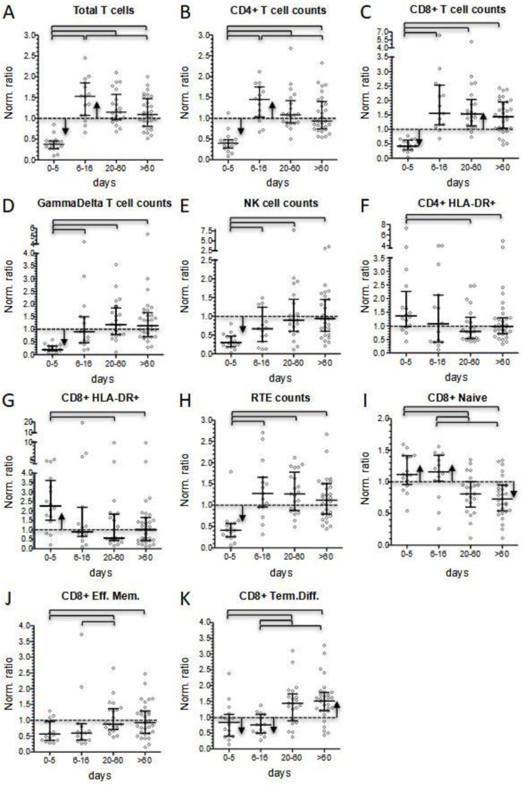
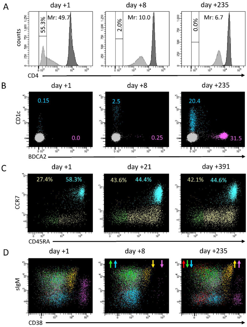
Results: MIS-C patients had elevated inflammatory markers, associated with T- and NK-cell lymphopenia, a profound depletion of dendritic cells, and altered monocyte phenotype at disease onset, while the subacute phase of the disease was characterized by a significant increase in T- and B-cell counts and a rapid decline in activated T cells and terminally differentiated B cells. Most of the immunological parameters returned to values close to the normal range during the remission phase (20-60 days after hospital admission). Nevertheless, we observed a significantly reduced ratio between recently generated and more differentiated CD8+ T- and B-cell subsets, which partially settled at longer-term follow-up determinations.

Conclusions: The characterization of lymphocyte distribution in different phases of MIS-C may help to understand the course of diseases that are associated with dysregulated immune responses and to calibrate prompt and targeted treatments.

Keywords: COVID; MIS-C; SARS-CoV-2; immunophenotype; lymphopenia; monocytopenia; multiparametric flow cytometry.


Conflict of interest statement

The authors declare no conflict of interest.

Figures

Figure 1
Figure 1
Patients’ biochemical and immunological parameters presented as longitudinal data (0–414 day from hospitalization) (left panels), and as comparison among data at hospital admission (Adm.), hospital discharge (Dis.), and follow-up (FU) (right panels). (A,B): C-reactive protein; (C,D): lymphocyte counts calculated as normalized ratios; (E,F): neutrophil absolute counts; (G,H): monocyte absolute counts. Bars of the scatter dot plots of panels (B,D,F,H) represent medians and interquartile range. Day-by-day analysis during hospitalization was performed by the Wilcoxon signed ranks test, while statistical analyses between groups of panels (B,D,F,H) were performed by the Mann–Whitney test. Horizontal segments indicate when p values were below 0.05.
Figure 2
Figure 2
Comparison of patients’ immunological parameters at four different time points according to days after hospital admission (0–5 and 6–16 days during hospitalization; 20–60 and >60 days at short- and long-term follow-up, respectively). (A): percentage of CD4- monocytes; (B) myeloid dendritic cells absolute counts; (C) plasmacytoid dendritic cells absolute counts; (D) basophils absolute counts. Bars of the scatter dot plots represent medians and interquartile range. Statistical analyses between groups were performed by the Mann–Whitney test. Horizontal bars indicate where p values were below 0.05.
Figure 3
Figure 3
Comparison of patients’ lymphocyte subsets values, calculated as normalized ratios, at four different time points according to days after hospital admission (0–5 and 6–16 days during hospitalization; 20–60 and >60 days at short and long-term follow-up, respectively). (A): Total T lymphocyte counts; (B) CD4+ T lymphocyte counts; (C) CD8+ T lymphocyte counts; (D) GammaDelta+ T lymphocyte counts; (E) NK cell counts; (F,G) percentage of activated CD4+ and CD8+ cells; (H) Recent Thymic Emigrants (RTE) counts; (IK) percentage of CD8+ cell subsets: naïve, effector, and terminally differentiated cells. Bars of the scatter dot plots represent medians and interquartile range. Statistical analyses between data for different time points were performed by the Mann–Whitney test. Horizontal bars among two groups indicate when p values were below 0.05. Pearson’s chi square test was performed to determine if data for each time point was significantly (p < 0.05) lower (down arrows) or higher (up arrows) from the normalized median (=1).
Figure 4
Figure 4
Comparison of patients’ B lymphocyte subset values, calculated as normalized ratios, at four different time points according to days after hospital admission (0–5 and 6–16 days during hospitalization; 20–60 and >60 days at short- and long-term follow-up, respectively). (A): Total B lymphocyte counts. Percentages of different B cell subsets: (B) recent bone marrow emigrants (RBE); (C) naïve; (D) switched memory; (E) IgM+ unswitched memory; (F) CD21low cells; (G) terminally differentiated. Bars of the scatter dot plots represent medians and interquartile range. Statistical analyses between data for different time points were performed by the Mann-Whitney test. Horizontal bars among two groups indicate when p values were below 0.05. Pearson’s chi square test was performed to determine if data for each time point was significantly (p < 0.05) lower (down arrows) or higher (up arrows) from the normalized median (=1).
Figure 5
Figure 5
Representative panel of longitudinal changes in immunological subsets in two MIS-C patients analyzed in three different phases of the disease. (A) CD4 expression on monocytes both measured as percentage of CD4- cells and MFI ratio (Mr) between T helper cells (gray histogram) and monocytes (white histogram); (B) pDC (pink) and mDC (blue) evaluation, according to BDCA2 and CD1c expression among CD45+CD4+CD14- gated cells; (C) evaluation of CD8+ T-cell maturation stages: naïve (blue), central memory (orange), effector memory (green), terminally differentiated (yellow); (D) evaluation of B cell subsets: RBE (orange), naïve (gray), switched memory (blue), unswitched memory (green), CD21low (red), terminally differentiated (pink). Arrows indicate when a subset significantly increased or decreased compared to the previous determination.

References

    1. Verdoni L., Mazza A., Gervasoni A., Martelli L., Ruggeri M., Ciuffreda M., Bonanomi E., D’Antiga L. An outbreak of severe Kawasaki-like disease at the Italian epicentre of the SARS-CoV-2 epidemic: An observational cohort study. Lancet. 2020;395:1771–1778. doi: 10.1016/S0140-6736(20)31103-X. - DOI - PMC - PubMed
    1. Whittaker E., Bamford A., Kenny J., Kaforou M., Jones C.E., Shah P., Ramnarayan P., Fraisse A., Miller O., Davies P., et al. Clinical Characteristics of 58 Children with a Pediatric Inflammatory Multisystem Syndrome Temporally Associated with SARS-CoV-2. JAMA. 2020;324:259–269. doi: 10.1001/jama.2020.10369. - DOI - PMC - PubMed
    1. Feldstein L.R., Rose E.B., Horwitz S.M., Collins J.P., Newhams M.M., Son M.B.F., Newburger J.W., Kleinman L.C., Heidemann S.M., Martin A.A., et al. Multisystem Inflammatory Syndrome in U.S. Children and Adolescents. N. Engl. J. Med. 2020;324:259–269. doi: 10.1056/NEJMoa2021680. - DOI - PMC - PubMed
    1. Cattalini M., Della Paolera S., Zunica F., Bracaglia C., Giangreco M., Verdoni L., Meini A., Sottile R., Caorsi R., Zuccotti G., et al. Defining Kawasaki disease and pediatric inflammatory multisystem syndrome-temporally associated to SARS-CoV-2 infection during SARS-CoV-2 epidemic in Italy: Results from a national, multicenter survey. Pediatr. Rheumatol. 2021;19:29. doi: 10.1186/s12969-021-00511-7. - DOI - PMC - PubMed
    1. Godfred-Cato S., Bryant B., Leung J., Oster M.E., Conklin L., Abrams J., Roguski K., Wallace B., Prezzato E., Koumans E.H., et al. COVID-19–Associated Multisystem Inflammatory Syndrome in Children—United States, March–July 2020. MMWR Morb. Mortal. Wkly. Rep. 2020;69:1074–1080. doi: 10.15585/mmwr.mm6932e2. - DOI - PMC - PubMed

LinkOut - more resources