

Mucoadhesive Rifampicin-Liposomes for the Treatment of Pulmonary Infection by Mycobacterium abscessus: Chitosan or ε-Poly-L-Lysine Decoration
- PMID: 37371504
- PMCID: PMC10296137
- DOI: 10.3390/biom13060924
Mucoadhesive Rifampicin-Liposomes for the Treatment of Pulmonary Infection by Mycobacterium abscessus: Chitosan or ε-Poly-L-Lysine Decoration
Abstract
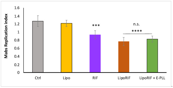
Mycobacterium abscessus (Mabs) is a dangerous non-tubercular mycobacterium responsible for severe pulmonary infections in immunologically vulnerable patients, due to its wide resistance to many different antibiotics which make its therapeutic management extremely difficult. Drug nanocarriers as liposomes may represent a promising delivery strategy against pulmonary Mabs infection, due to the possibility to be aerosolically administrated and to tune their properties in order to increase nebulization resistance and retainment of encapsulated drug. In fact, liposome surface can be modified by decoration with mucoadhesive polymers to enhance its stability, mucus penetration and prolong its residence time in the lung. The aim of this work is to employ Chitosan or ε-poly-L-lysine decoration for improving the properties of a novel liposomes composed by hydrogenated phosphatidyl-choline from soybean (HSPC) and anionic 1,2-Dipalmitoyl-sn-glycero-3-phosphorylglycerol sodium salt (DPPG) able to entrap Rifampicin. A deep physicochemical characterization of polymer-decorated liposomes shows that both polymers improve mucoadhesion without affecting liposome features and Rifampicin entrapment efficiency. Therapeutic activity on Mabs-infected macrophages demonstrates an effective antibacterial effect of ε-poly-L-lysine liposomes with respect to chitosan-decorated ones. Altogether, these results suggest a possible use of ε-PLL liposomes to improve antibiotic delivery in the lung.
Keywords: Chitosan; Mycobacterium abscessus; Rifampicin; liposomes; mucoadhesion; polymer decoration; ε-poly-L-lysine.
Conflict of interest statement
The authors declare no conflict of interest.
Figures


Similar articles
-
Rifampicin-Liposomes for Mycobacterium abscessus Infection Treatment: Intracellular Uptake and Antibacterial Activity Evaluation.Pharmaceutics. 2021 Jul 13;13(7):1070. doi: 10.3390/pharmaceutics13071070. Pharmaceutics. 2021. PMID: 34371761 Free PMC article.
-
Combined Host- and Pathogen-Directed Therapy for the Control of Mycobacterium abscessus Infection.Microbiol Spectr. 2022 Feb 23;10(1):e0254621. doi: 10.1128/spectrum.02546-21. Epub 2022 Jan 26. Microbiol Spectr. 2022. PMID: 35080463 Free PMC article.
-
Liposomes coated with chitosan-xanthan gum (chitosomes) as potential carriers for pulmonary delivery of rifampicin.J Pharm Sci. 2012 Feb;101(2):566-75. doi: 10.1002/jps.22775. Epub 2011 Oct 13. J Pharm Sci. 2012. PMID: 21997465
-
Lipid-polymer hybrid nanoparticles as a new generation therapeutic delivery platform: a review.Eur J Pharm Biopharm. 2013 Nov;85(3 Pt A):427-43. doi: 10.1016/j.ejpb.2013.07.002. Epub 2013 Jul 17. Eur J Pharm Biopharm. 2013. PMID: 23872180 Review.
-
Overcoming resistance: Chitosan-modified liposomes as targeted drug carriers in the fight against multidrug resistant bacteria-a review.Int J Biol Macromol. 2024 Oct;278(Pt 4):135022. doi: 10.1016/j.ijbiomac.2024.135022. Epub 2024 Aug 23. Int J Biol Macromol. 2024. PMID: 39182895 Review.
Cited by
-
Chitosan-Based Nanoformulations: Preclinical Investigations, Theranostic Advancements, and Clinical Trial Prospects for Targeting Diverse Pathologies.AAPS PharmSciTech. 2024 Nov 5;25(8):263. doi: 10.1208/s12249-024-02948-x. AAPS PharmSciTech. 2024. PMID: 39500815 Review.
-
Application of chitosan-based drug delivery systems in the treatment of bacterial diseases: a review.Drug Deliv. 2025 Dec;32(1):2514140. doi: 10.1080/10717544.2025.2514140. Epub 2025 Jun 10. Drug Deliv. 2025. PMID: 40491201 Free PMC article. Review.
-
Liposome-Encapsulated Antibiotics for the Therapy of Mycobacterial Infections.Antibiotics (Basel). 2025 Jul 20;14(7):728. doi: 10.3390/antibiotics14070728. Antibiotics (Basel). 2025. PMID: 40724029 Free PMC article. Review.
-
Enhancing Human Health Through Nutrient and Bioactive Compound Recovery from Agri-Food By-Products: A Decade of Progress.Nutrients. 2025 Jul 31;17(15):2528. doi: 10.3390/nu17152528. Nutrients. 2025. PMID: 40806112 Free PMC article. Review.
-
Combined System for the Simultaneous Delivery of Levofloxacin and Rifampicin: Structural and Functional Properties and Antibacterial Activity.J Funct Biomater. 2023 Jul 20;14(7):381. doi: 10.3390/jfb14070381. J Funct Biomater. 2023. PMID: 37504876 Free PMC article.
References
-
- Marianecci C., Marzio L.D., Rinaldi F., Carafa M., Alhaique F. Pulmonary Delivery: Innovative Approaches and Perspectives. J. Biomater. Nanobiotechnol. 2011;2:567. doi: 10.4236/jbnb.2011.225068. - DOI
Publication types
MeSH terms
Substances
LinkOut - more resources
Full Text Sources
