. 2023 May 24;11(6):1517.
doi: 10.3390/biomedicines11061517.

CCR4 Blockade Diminishes Intratumoral Macrophage Recruitment and Augments Survival of Syngeneic Pancreatic Cancer-Bearing Mice

Affiliations

CCR4 Blockade Diminishes Intratumoral Macrophage Recruitment and Augments Survival of Syngeneic Pancreatic Cancer-Bearing Mice

Aydar Khabipov et al. Biomedicines. .

Abstract

Pancreatic cancer is known for its tumor microenvironment (TME), which is rich in stromal and immune cells supporting cancer growth and therapy resistance. In particular, tumor-associated macrophages (TAMs) are known for their angiogenesis- and metastasis-promoting properties, which lead to the failure of conventional therapies for pancreatic cancer. Hence, treatment options targeting TAMs are needed. The C-C chemokine receptor type 4 (CCR4) is critical for immune cell recruitment into the TME, and in this paper we explore the effects of its genetic or immunotherapeutic blockade in pancreatic-cancer-bearing mice. Murine PDA6606 pancreatic cancer cells and murine peritoneal macrophages were used for in vitro migration assays. In vivo, a syngeneic, orthotropic pancreatic cancer model was established. Tumor growth and survival were monitored under prophylactic and therapeutic application of a CCR4 antagonist (AF-399/420/18025) in wildtype (CCR4wt) and CCR4-knockout (CCR4-/-) mice. Immune infiltration was monitored in tumor tissue sections and via flow cytometry of lysed tumors. PDA6606 cells induced less migration in CCR4-/- than in CCR4wt macrophages in vitro. Pancreatic TAM infiltration was higher, and survival was reduced in CCR4wt mice compared to CCR4-/- mice. Antagonizing CCR4 in wildtype mice revealed similar results as in CCR4-/- mice without antagonization. Prophylactic CCR4 antagonist application in wildtype mice was more efficient than therapeutic antagonization. CCR4 seems to be critically involved in TAM generation and tumor progression in pancreatic cancer. CCR4 blockade may help prolong the relapse-free period after curative surgery in pancreatic cancer and improve prognosis.

Keywords: CCL17; CCL22; M2 macrophages; TAMs; migration; tumor-associated macrophages.


Conflict of interest statement

The authors declare no conflict of interest.

Figures

Figure 1
Figure 1
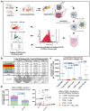
Effects of CCR4 knockout/CCR4 antagonist on tumor growth. (a) Settings of in vivo experiments: pancreatic carcinoma (PC) cells were implanted in the pancreas of wildtype mice as controls (C-group)* and CCR4−/− mice (KO-group) the CCR4 antagonist (AF399/420/18025) was given to wildtype mice 2 days before PC implantation (P-group)*** or 2 weeks after tumor implantation (T-group); PC was implanted in wildtype mice as a control, DMSO was given instead of AF399/420/18025 in the T- and P-groups as a control, volumes were measured via MRI after 3 and 5 weeks in the P- and T-groups or after 3 and 6 weeks in the KO group. (b) Photograph of 7-tesla-PET magnetic resonance imager (i) and preparation of mice for imaging (ii). (c) MRI images for volume measurement in the sagittal (left) and transversal (right) dimensions; tumors are marked in blue (control) (i), yellow (P-group) with prophylactic CCR4-blockade (ii), and red (T-group) with therapeutic CCR4 blockade (iii). (df) Tumor volume in the CCR4−/− group (KO) after 3 and 6 weeks (d), tumor volume in the CCR4 block (prophylactic) group (P) after 3 and 5 weeks (e), and tumor volume in the CCR4 block (therapeutic) group (T) after 3 and 5 weeks (f). The significance levels are indicated as follows: * = p < 0.05, *** = p < 0.001. n.s. = not significant.
Figure 2
Figure 2
Survival dynamics of CCR4-blocked mice with pancreatic cancer. (a) Survival of CCR4-knockout mice with pancreatic cancer (PC)(orange) vs. controls (wildtype with PC). (b) CCR4-blocked mice with PC (prophylactic; orange) vs. controls (wildtype with PC; blue). (c) CCR4-blocked mice with PC (therapeutic; orange) vs. controls (wildtype mice with PC). The x-axis shows days.
Figure 3
Figure 3
Quantifying intratumoral immune cells via flow cytometry. (a) Experimental settings: tumors were implanted in wildtype mice and CCR4−/− mice as described, explantation was performed after 5 weeks, tumors were minced manually with scissors, collagenase, and DNase were used for tumor lysis, single-cell isolation was performed through 100 µm and 40 µm pore filters; intratumoral immune cells were detected via CD45 and subclassified to macrophages, B lymphocytes, CD4+ T cells, CD8+ T cells, and Tregs via antibody labeling and flow cytometry. (b) Composition of immune cells concerning all tumor cells (or immune cells only) in wildtype mice and CCR4−/− mice (tabular). (c) Composition of intratumoral immune cells in wildtype mice. (d) CCR4 knockout effect on immune cell infiltration (left) and subtypes (right). The significance levels are indicated as follows: * = p < 0.05, ** = p < 0.01, *** = p < 0.001. n.s. = not significant.
Figure 4
Figure 4
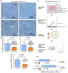
CCR4 knockout/antagonist effects on macrophage migration and infiltration. (a) Immunohistological tissue stains of in vivo tumors with mac receptor F4/80 show reduced infiltration of macrophages (mac) in the tumor after prophylactic CCR4 blockade and therapeutic treatment with CCR4 blockade compared to controls; CCR4−/− tumors present similar results (histological stain not shown). (b) Quantification of F4/80-positive cells (alias macrophages), showing significantly reduced infiltration in CCR4-KO tumors. (iii); tumors of CCR4-antagonized mice also presented less macrophage infiltration in both the prophylactic and therapeutic groups (i,ii). (c) Workflow protocol of migration assay using Boyden chamber, murine intraperitoneal MCS-F-stimulated macrophages were placed in Matrigel and seeded in inserts, prior to the well bottoms being filled with PDA6606 supernatants as a migration stimulus; after 12 h of incubation, the insert was removed, and migrated cells were disengaged from the monolayer and collected for quantification via FACS. (d) In vitro migration of peritoneal macrophages of CCR4-KO-mice was reduced compared to wildtype macrophages. PC (peritoneal carcinomatosis)-cell-supernatant-stimulated macrophages presented increased migration compared to non-stimulated macrophages in both the CCR4 and wildtype groups. The significance levels are indicated as follows: * = p < 0.05, ** = p < 0.01.
Figure 5
Figure 5
Principle of the therapeutic concept of a CCR4 antagonist in pancreatic cancer. (a) Monocyte recruitment via CCR4 ligands CCL17/CCL22 leads to TAM development and peritumoral angiogenesis. (b) Monocyte recruitment is inhibited by blocking CCR4 on monocytes. Consequently, monocyte infiltration and TAM development are decreased. Hence, angiogenesis and tumor progress are reduced.

References

    1. Sokol C.L., Luster A.D. The Chemokine System in Innate Immunity. Cold Spring Harb. Perspect. Biol. 2015;7:a016303. doi: 10.1101/cshperspect.a016303. - DOI - PMC - PubMed
    1. Ness T.L., Ewing J.L., Hogaboam C.M., Kunkel S.L. Ccr4 Is a Key Modulator of Innate Immune Responses. J. Immunol. 2006;177:7531–7539. doi: 10.4049/jimmunol.177.11.7531. - DOI - PubMed
    1. Al-Banna N.A., Vaci M., Slauenwhite D., Johnston B., Issekutz T.B. Ccr4 and Cxcr3 Play Different Roles in the Migration of T Cells to Inflammation in Skin, Arthritic Joints, and Lymph Nodes. Eur. J. Immunol. 2014;44:1633–1643. doi: 10.1002/eji.201343995. - DOI - PubMed
    1. Morimoto Y., Bian Y., Gao P., Yashiro-Ohtani Y., Zhou X.Y., Ono S., Nakahara H., Kogo M., Hamaoka T., Fujiwara H. Induction of Surface Ccr4 and Its Functionality in Mouse Th2 Cells Is Regulated Differently During Th2 Development. J. Leukoc. Biol. 2005;78:753–761. doi: 10.1189/jlb.0305139. - DOI - PubMed
    1. Wang Z., Louras N.J., Lellouch A.G., Pratts S.G., Zhang H., Wang H., Huang C.A., Cetrulo C.L., Jr., Madsen J.C., Sachs D.H., et al. Dosing Optimization of Ccr4 Immunotoxin for Improved Depletion of Ccr4(+) Treg in Nonhuman Primates. Mol. Oncol. 2018;12:1374–1382. doi: 10.1002/1878-0261.12331. - DOI - PMC - PubMed

LinkOut - more resources