. 2023 May 26;11(6):1544.
doi: 10.3390/biomedicines11061544.

Interaction of Drug-Sensitive and -Resistant Human Melanoma Cells with HUVEC Cells: A Label-Free Cell-Based Impedance Study

Affiliations

Interaction of Drug-Sensitive and -Resistant Human Melanoma Cells with HUVEC Cells: A Label-Free Cell-Based Impedance Study

Giuseppina Bozzuto et al. Biomedicines. .

Abstract

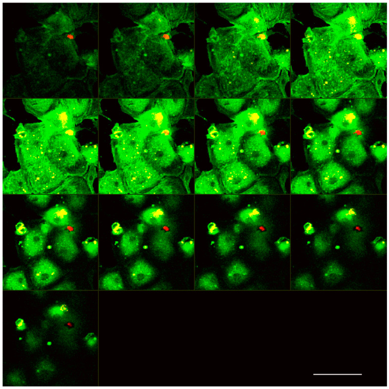
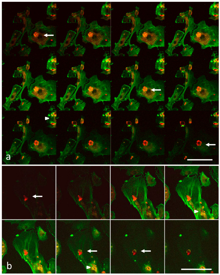
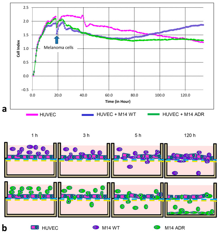
Cancer cell extravasation is a crucial step in cancer metastasis. However, many of the mechanisms involved in this process are only now being elucidated. Thus, in the present study we analysed the trans-endothelial invasion of melanoma cells by a high throughput label-free cell impedance assay applied to transwell chamber invasion assay. This technique monitors and quantifies in real-time the invasion of endothelial cells by malignant tumour cells, for a long time, avoiding artefacts due to preparation of the end point measurements. Results obtained by impedance analysis were compared with endpoint measurements. In this study, we used human melanoma M14 wild type (WT) cells and their drug resistant counterparts, M14 multidrug resistant (ADR) melanoma cells, selected by prolonged exposure to doxorubicin (DOX). Tumour cells were co-cultured with monolayers of human umbilical vein endothelial cells (HUVEC). Results herein reported demonstrated that: (i) the trans-endothelial migration of resistant melanoma cells was faster than sensitive ones; (ii) the endothelial cells appeared to be strongly affected by the transmigration of melanoma cells which showed the ability to degrade their cytoplasm; (iii) resistant cells preferentially adopted the transcellular invasion vs. the paracellular one; (iv) the endothelial damage mediated by tumour metalloproteinases seemed to be reversible.

Keywords: cancer cell extravasation; human melanoma cells; label-free cell impedance assay; light and electron microscopy; multidrug resistance.


Conflict of interest statement

The authors declare no conflict of interest.

Figures

Scheme 1
Scheme 1
Schematic representation of (a) CIM-Plate 16 well and (b) impedance-based measuring theory.
Figure 1
Figure 1
Laser scanning confocal microscopy. Serial optical sections (from the top to the bottom) of the M14WT/HUVEC co-cultures after 1 h of interaction: the red signal comes from tagged melanoma cells, the green signal from fluorescein actin of HUVEC cells, and the yellow signal reveals the interaction of tumour cells with endothelial cells. Bar: 100 µm.
Figure 2
Figure 2
Laser scanning confocal microscopy. (a,b) Serial optical sections from the top to the bottom M14WT/HUVEC co-cultures. After 3 h of interaction, sensitive melanoma cells appear to infiltrate both between adjacent HUVEC cells (intercellular invasion) (arrows) and within isolated cells (arrow heads) (transcellular invasion). Bar: 100 µm.
Figure 3
Figure 3
Laser scanning confocal microscopy. Serial optical sections from the top to the bottom of M14WT/HUVEC cell co-cultures after 5 h of interaction. (a) Actin-labelled HUVEC cell. (b) Merged signal of clustered M14 WT cells (red signal, arrow heads) undergoing transcellular invasion. HUVEC F-actin is assembled into a ring-like array around the circumference of the invasion pore (green signal, arrow) that seems to encapsulate the invading cancer cells. The yellow signal represents the co-localisation of HUVEC and melanoma cell structures. Bar: 100 µm.
Figure 4
Figure 4
Laser scanning confocal microscopy. Serial optical sections of M14 ADR/HUVEC cell co-cultures, after 3 h of interaction. (a) XY sections. (b) XZ sections. A high number of melanoma cells (red signal) interacting with endothelial cells (green signal) is visible. Bar: 100 µm.
Figure 5
Figure 5
Laser scanning confocal microscopy. Serial optical sections of M14 ADR/HUVEC cell co-cultures, after five hours of interaction. (a) Resistant melanoma cells were able to complete the trans-endothelial migration. (b) Also in these samples, a ring-like actin array (yellow, arrow) around the circumference of the invasion pore is visible. Bar: 100 µm.
Figure 6
Figure 6
Scanning electron microscopy observations of M14WT/HUVEC co-cultures after 1 h of interaction. (a) HUVEC cell (asterisk), melanoma cells (arrow). (b,c) Tumour cells (arrow) show the surface covered by randomly distributed microvilli and blebs. Contact regions also show thin microvilli arising from the underlying endothelial cells (arrow heads). Insert: Image at low magnification of the co-culture. (a,c) Bar: 20 µm. (c) Bar: 10 µm.
Figure 7
Figure 7
Scanning electron microscopy observations of M14 ADR/HUVEC co-cultures after 1 h of interaction. (a) HUVEC cell (asterisk) displayed several ruptures and holes of the cytoplasm. (b,c) Melanoma cell (arrow) prolongs cellular protrusions (leading edge) rich of vesicles that pierces the cytoplasm of endothelial cell. Insert: Image at low magnification of the co-culture. (a) Bar: 50 µm. (b,c) Bar: 1 µm.
Figure 8
Figure 8
Scanning electron microscopy observations of M14WT/HUVEC co-cultures after 3 h of interaction. (a) HUVEC cells (asterisks), melanoma cells (arrows). (b) The involvement of endothelial cells in the interaction with tumour cells is confirmed. (c) Melanoma cell penetrated in a endothelial cell. Insert: Image at low magnification of the co-culture. (a) Bar: 50 µm. (b,c) Bar: 20 µm.
Figure 9
Figure 9
Scanning electron microscopy observations of M14 ADR/HUVEC co-cultures after 3 h of interaction. (a) HUVEC cell (asterisk) interacting with resistant melanoma cells displayed a dramatically altered morphology with several ruptures and holes of the cytoplasm. (b) Melanoma cell penetrating HUVEC cell. Arrows in (a,b) indicate melanoma cells. (c) Detail of image (b) at higher magnification. Top right insert: Image at low magnification of the co-culture. (a) Bar: 50 µm. (b) Bar: 10 µm. (c) Bar: 5 µm.
Figure 10
Figure 10
Scanning electron microscopy observations of M14WT/HUVEC co-cultures after 5 h of interaction. (a,b) Endothelial cells still appeared damaged. (b) A melanoma cell passing through the cytoplasm of a HUVEC cell is visible. (a) Bar: 100 µm. (b) Bar: 50 µm. Insert Bar: 10 µm.
Figure 11
Figure 11
Scanning electron microscopy observations of M14ADR/HUVEC co-cultures after 5 h of interaction. (a,b) As resistant melanoma cells came into contact with the substrate, they spread on the surface and adopted a fibroblastic morphology, becoming sandwiched. (b) When the migration of tumour cells was completed, endothelial cells recovered their damaged morphology. (a) Bar: 100 µm. (b) Bar: 50 µm.
Figure 12
Figure 12
Transmission electron microscopy. Melanoma cells (MC) adhering and invading endothelial cells (EC). (ac) It is possible to observe a melanoma cell completely immersed in the cytoplasm of HUVEC cell. (df) Adhering tumour cells degrading the cytoplasm of endothelial cells (asterisk). (ad) Bar: 10 µm. (e,f) Bar: 5 µm.
Figure 13
Figure 13
Zymography. Following the interaction with HUVEC cells, both drug-sensitive and resistant melanoma cells were able to release MMP9 gelatinase (arrow). However, the amount released by resistant cells was higher than that of sensitive ones, after both 5 and 24 h of interaction. Conditioned medium from BHK-21 (BHK CM) cells was used as a positive control.
Figure 14
Figure 14
Electrical impedance assay. (a) Cell index of HUVEC/melanoma cells continuously monitored by xCELLigence RTCA DP system. Arrow indicates the addition of melanoma cells to HUVEC monolayer. (b) Proposed model of M14 WT and M14 ADR transmigration through the endothelial monolayer.

References

    1. Liotta L.A., Rao C.N., Barsky S.H. Tumor invasion and the extracellular matrix. Lab. Investig. 1983;49:636–649. - PubMed
    1. Orr F.W., Wang H.H., Lafrenie R.M., Scherbarth S., Nance D.M. Interactions between cancer cells and the endothelium in metastasis. J. Pathol. 2000;190:310–329. doi: 10.1002/(SICI)1096-9896(200002)190:3<310::AID-PATH525>3.0.CO;2-P. - DOI - PubMed
    1. Haier J., Korb T., Hotz B., Spiegel H.-U., Senninger N. An intravital model to monitor steps of metastatic tumor cell adhesion within the epatic microcirculation. J. Gastrointest. Surg. 2003;7:507–515. doi: 10.1016/S1091-255X(03)00023-4. - DOI - PubMed
    1. Salminen A.T., Allahyari Z., Gholizadeh S., McCloskey M.C., Ajalik R., Cottle R.N., Gaborski T.R., McGrath J.L. In vitro Studies of Transendothelial Migration for Biological and Drug Discovery. Front. Med. Technol. 2020;2:600616. doi: 10.3389/fmedt.2020.600616. - DOI - PMC - PubMed
    1. Nicolson G.L. Metastatic tumor cell interactions with endothelium, basement membrane and tissue. Curr. Opin. Cell Biol. 1989;1:1009–1019. doi: 10.1016/0955-0674(89)90073-2. - DOI - PubMed