. 2023 May 27;11(6):1559.
doi: 10.3390/biomedicines11061559.

Circulating Angiogenic Factors and Ischemic Diabetic Foot Syndrome Advancement-A Pilot Study

Affiliations

Circulating Angiogenic Factors and Ischemic Diabetic Foot Syndrome Advancement-A Pilot Study

Martyna Schönborn et al. Biomedicines. .

Abstract

Despite clear evidence of inadequate angiogenesis in ischemic diabetic foot syndrome (DFS) pathogenesis, angiogenic factor level changes in patients with ischemic DFS remain inconsistent. This study aimed to assess circulating angiogenic factors concerning ischemic DFS advancement and describe their relationships with patients' clinical characteristics, microvascular parameters, and diabetic control. The study included 41 patients with ischemic DFS (67.3 (8.84) years; 82.9% males). Angiogenic processes were assessed by identifying circulating concentrations of five pro- and two anti-angiogenic factors. We found that penetrating ulcers were related to a significantly higher FGF-2 level (8.86 (5.29) vs. 5.23 (4.17) pg/mL, p = 0.02). Moreover, plasma FGF-2 showed a significant correlation with the SINBAD score (r = 0.32, p = 0.04), platelet count (r = 0.43, p < 0.01), white cell count (r = 0.42, p < 0.01), and age (r = -0.35, p = 0.03). We did not observe any significant linear relationship between the studied biomarkers and microcirculatory parameters, nor for glycemic control. In a univariate analysis using logistic regression, an increase in plasma FGF-2 was tied to greater odds of high-grade ulcers (OR 1.16; 95% CI 1.02-1.38, p = 0.043). This suggests that circulating FGF-2 may serve as a potential biomarker for predicting DFU advancement and progression. It is necessary to conduct further studies with follow-up observations to confirm this hypothesis.

Keywords: angiogenesis; angiogenic factors; diabetic foot syndrome; microcirculation; peripheral arterial disease.


Conflict of interest statement

The authors declare no conflict of interest.

Figures

Figure 1
Figure 1
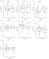
Boxplots with jittered data illustrating the relationships between plasma FGF-2 and (A) coronary artery disease, (B) statin use, (C) wound infection, (D) smoking status (yes—active or past smoker), and (E) ulcer depth FGF-2—fibroblast growth factor 2.
Figure 2
Figure 2
Scatterplots with a linear fit illustrating the relationships between plasma FGF-2 and (A) age, (B) BMI, (C) WBC, (D) total cholesterol, (E) PLT, (F) LDF, and (G) tcpO2. FGF-2—fibroblast growth factor 2, BMI—body mass index, WBC—white blood cells count, PLT—platelet count, LDF—laser Doppler flowmetry, and tcpO2—transcutaneous oximetry test.
Figure 3
Figure 3
Boxplots with jittered data illustrating the relationships between plasma VEGF-A and (A) coronary artery disease, (B) statin use, (C) wound infection, (D) smoking status (yes—active or past smoker), and (E) ulcer depth VEGF-A—vascular endothelial growth factor A.
Figure 4
Figure 4
Scatterplots with a linear fit illustrating the relationships between plasma VEGF-A and (A) age, (B) BMI, (C) WBC, (D) total cholesterol, (E) PLT, (F) LDF, and (G) tcpO2. VEGF-A—vascular endothelial growth factor A, BMI—body mass index, WBC—white blood cells count, PLT—platelet count, LDF—laser Doppler flowmetry, and tcpO2—transcutaneous oximetry test.
Figure 5
Figure 5
Boxplots with jittered data illustrating the relationships between plasma VEGF-R2 and (A) coronary artery disease, (B) statin use, (C) wound infection, (D) smoking status (yes—active or past smoker), and (E) ulcer depth VEGF-R2—vascular endothelial growth factor receptor 2.
Figure 6
Figure 6
Scatterplots with a linear fit illustrating the relationships between plasma VEGF-R2 and (A) age, (B) BMI, (C) WBC, (D) total cholesterol, (E) PLT, (F) LDF, and (G) tcpO2. VEGF-R2—vascular endothelial growth factor receptor 2, BMI—body mass index, WBC—white blood cells count, PLT—platelet count, LDF—laser Doppler flowmetry, and tcpO2—transcutaneous oximetry test.
Figure 7
Figure 7
Boxplots with jittered data illustrating the relationships between plasma PlGF and (A) coronary artery disease, (B) statin use, (C) wound infection, (D) smoking status (yes—active or past smoker), and (E) ulcer depth PlGF—placental growth factor.
Figure 8
Figure 8
Scatterplots with a linear fit illustrating the relationships between plasma PlGF and (A) age, (B) BMI, (C) WBC, (D) total cholesterol, (E) PLT, (F) LDF, and (G) tcpO2. PlGF—placental growth factor, BMI—body mass index, WBC—white blood cells count, PLT—platelet count, LDF—laser Doppler flowmetry, and tcpO2—transcutaneous oximetry test.
Figure 9
Figure 9
Boxplots with jittered data illustrating the relationships between plasma PDGF-BB and (A) coronary artery disease, (B) statin use, (C) wound infection, (D) smoking status (yes—active or past smoker), and (E) ulcer depth PDGF-BB—platelet-derived growth factor-BB.
Figure 10
Figure 10
Scatterplots with a linear fit illustrating the relationships between plasma PDGF-BB and (A) age, (B) BMI, (C) WBC, (D) total cholesterol, (E) PLT, (F) LDF, and (G) tcpO2. PDGF-BB—platelet-derived growth factor-BB, BMI—body mass index, WBC—white blood cells count, PLT—platelet count, LDF—laser Doppler flowmetry, and tcpO2—transcutaneous oximetry test.
Figure 11
Figure 11
Boxplots with jittered data illustrating the relationships between plasma PEDF and (A) coronary artery disease, (B) statin use, (C) wound infection, (D) smoking status (yes—active or past smoker), and (E) ulcer depth PEDF—pigment epithelium-derived factor.
Figure 12
Figure 12
Scatterplots with a linear fit illustrating the relationships between plasma PEDF and (A) age, (B) BMI, (C) WBC, (D) total cholesterol, (E) PLT, (F) LDF, and (G) tcpO2. PEDF—pigment epithelium-derived factor, BMI—body mass index, WBC—white blood cells count, PLT—platelet count, LDF—laser Doppler flowmetry, and tcpO2—transcutaneous oximetry test.
Figure 13
Figure 13
Boxplots with jittered data illustrating the relationships between plasma ANG1 and (A) coronary artery disease, (B) statin use, (C) wound infection, (D) smoking status (yes—active or past smoker), and (E) ulcer depth ANG1—angiopoietin-1.
Figure 14
Figure 14
Scatterplots with a linear fit illustrating the relationships between plasma ANG1 and (A) age, (B) BMI, (C) WBC, (D) total cholesterol, (E) PLT, (F) LDF, and (G) tcpO2. ANG-1—angiopoietin-1, BMI—body mass index, WBC—white blood cells count, PLT—platelet count, LDF—laser Doppler flowmetry, and tcpO2—transcutaneous oximetry test.
Figure 15
Figure 15
Box and jitter plots illustrating the relationships between (A) VEGFR2, (B) VEGF-A, (C) PIGF, (D) PDGF-BB, (E) FGF-2, (F) PEDF, and (G) ANG-1 and glycemic control (HBA1c cut-off at 8%). VEGF-A—vascular endothelial growth factor A, VEGF-R2—vascular endothelial growth factor receptor 2, FGF-2—fibroblast growth factor 2, PlGF—placental growth factor, PDGF-BB—platelet-derived growth factor-BB, PEDF—pigment epithelium-derived factor, and ANG-1—angiopoietin-1.
Figure 16
Figure 16
A diagram illustrating the potential double pathway of FGF-2 action on impaired wound healing in the course of DFS. ↑ increased, ↓ decreased.

References

    1. Zhang P., Lu J., Jing Y., Tang S., Zhu D., Bi Y. Global epidemiology of diabetic foot ulceration: A systematic review and meta-analysis. Ann. Med. 2017;49:106–116. doi: 10.1080/07853890.2016.1231932. - DOI - PubMed
    1. Armstrong D.G., Boulton A.J.M., Bus S.A. Diabetic Foot Ulcers and Their Recurrence. N. Engl. J. Med. 2017;376:2367–2375. doi: 10.1056/NEJMra1615439. - DOI - PubMed
    1. van Netten J.J., Bus S.A., Apelqvist J., Lipsky B.A., Hinchliffe R.J., Game F., Rayman G., Lazzarini P.A., Forsythe R.O., Peters E.J.G., et al. Definitions and criteria for diabetic foot disease. Diabetes Metab. Res. Rev. 2020;36:e3268. doi: 10.1002/dmrr.3268. - DOI - PubMed
    1. Bandyk D.F. The diabetic foot: Pathophysiology, evaluation, and treatment. Semin. Vasc. Surg. 2018;31:43–48. doi: 10.1053/j.semvascsurg.2019.02.001. - DOI - PubMed
    1. Spiliopoulos S., Festas G., Paraskevopoulos I., Mariappan M., Brountzos E. Overcoming ischemia in the diabetic foot: Minimally invasive treatment options. World J. Diabetes. 2021;12:2011–2026. doi: 10.4239/wjd.v12.i12.2011. - DOI - PMC - PubMed

LinkOut - more resources