Review
. 2023 Jun 9;11(6):1674.
doi: 10.3390/biomedicines11061674.

Pre- and Post-Transcriptional Control of HBV Gene Expression: The Road Traveled towards the New Paradigm of HBx, Its Isoforms, and Their Diverse Functions

Affiliations
Review

Pre- and Post-Transcriptional Control of HBV Gene Expression: The Road Traveled towards the New Paradigm of HBx, Its Isoforms, and Their Diverse Functions

Rodrigo A Villanueva et al. Biomedicines. .

Abstract

Hepatitis B virus (HBV) is an enveloped DNA human virus belonging to the Hepadnaviridae family. Perhaps its main distinguishable characteristic is the replication of its genome through a reverse transcription process. The HBV circular genome encodes only four overlapping reading frames, encoding for the main canonical proteins named core, P, surface, and X (or HBx protein). However, pre- and post-transcriptional gene regulation diversifies the full HBV proteome into diverse isoform proteins. In line with this, hepatitis B virus X protein (HBx) is a viral multifunctional and regulatory protein of 16.5 kDa, whose canonical reading frame presents two phylogenetically conserved internal in-frame translational initiation codons, and which results as well in the expression of two divergent N-terminal smaller isoforms of 8.6 and 5.8 kDa, during translation. The canonical HBx, as well as the smaller isoform proteins, displays different roles during viral replication and subcellular localizations. In this article, we reviewed the different mechanisms of pre- and post-transcriptional regulation of protein expression that take place during viral replication. We also investigated all the past and recent evidence about HBV HBx gene regulation and its divergent N-terminal isoform proteins. Evidence has been collected for over 30 years. The accumulated evidence simply strengthens the concept of a new paradigm of the canonical HBx, and its smaller divergent N-terminal isoform proteins, not only during viral replication, but also throughout cell pathogenesis.

Keywords: HBx; hepatitis B virus; hepatitis B virus X protein; isoform protein; mRNA; pre- and post-transcriptional control; viral regulatory protein; viral replication.


Conflict of interest statement

The authors declare no conflict of interest.

Figures

Figure 5
Figure 5
HBV HBx gene expression is controlled by both alternative translation and transcription initiation. At the top, HBV HBx DNA where the X promoter and the embedded intragenic promoter are shown. TSSs for HBV nucleotides 1210 and 1523 are shown [132] as well as the predicted HBx mRNAs, corresponding to XI and XII promoter transcription, in red (Supplementary Table S1). Below, the canonical HBx protein as well as the two smaller, divergent N-terminal isoform proteins: middle and small (mini) HBx [142].
Figure 1
Figure 1
Hepatitis B circular genome. The HBV genome is a partially double-stranded, relaxed, and circular DNA of about 3.2 kb. The genome contains four overlapping reading frames encoding for the viral envelope or surface (pre-S1/pre-S2/S, red arrow), core (pre-core/core) (green arrow), polymerase (blue arrow), and HBx protein (yellow arrow). As shown, the DNA genome contains two direct repeats (DR1 and DR2), two enhancers (EnhI, EnhII), and four promoter regions that regulate protein expression. Viral P protein is covalently bound to the genome (blue) and an RNA primer is bound to the negative strand (gray).
Figure 2
Figure 2
HBV mRNAs and their coding capacity. (A) pg-RNA, 3.4 kb. pg-RNA is organized in a bi-cistronic arrangement where the first AUG is encoding for core, and downstream, in a different reading frame, the AUG codes for the P protein. Core and P proteins are shown with their main domain organizations. (B) preC-RNA, 3.5 kb. preC protein starts translation upstream to the core’s AUG, but in the same reading frame. Sites for cleavage events (ER and Golgi apparatus) on the immature protein are shown. As a result, HBeAg is a secreted protein. (C) preS1-RNA, 2.4 kb. At the top, HBV DNA is shown with the relative locations of S1 and S2 promoters. preS1-RNA is predicted to encode the canonical LHBs, although other AUGs are present in the same reading frame (see the text). LHBs is shown with its domains preS1, preS2, and S. Both preS1 and S domains bear PTMs as shown. (D) S-RNA, 2.1 kb. S-RNA is predicted to encode both preS2 and S domains where MHBs and SHBs are produced.
Figure 3
Figure 3
Hepatitis B-spliced protein, HBSP. HBSP is produced from a single splicing event, and it is encoded by the 2.2 kb sp1 RNA molecule of the HBV pg-mRNA transcript. Both core and P reading frames are shown. SD, splice donor site, SA, splice acceptor site, and intron, are shown, located within the P reading frame. During translation initiation, the 47 first residues of P protein are translated, and then the junction changes the reading frame, and so the sequence of the C-terminal half of this protein is unique.
Figure 4
Figure 4
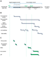
Domain organization of the canonical HBx protein and interacting partners. (A) HBx protein is organized into two distinct functional domains. The N-terminal negative regulatory domain (residues 1–50) is composed of a highly conserved N-terminal region and a Ser/Pro-rich region as indicated. The C-terminal region contains the crucial transactivation domain covering the AUG2 (Met79) and AUG3 (Met105). Both the H-box alpha-helix and the BH3-like motif are shown. (B) Mapped location of HBx functional regions. Regions of transrepressor activity, transactivation domain, signal transduction, nuclear transactivation, putative mitochondrial localization, and protein stability are shown. (C) Mapped interacting partners. Regions of interactions with Prdx1, 14-3-3, DDB1, Bcl-2, and p53 proteins are shown.
Figure 6
Figure 6
Scheme of the predicted residue sequence of the HBwx protein. Canonical HBx protein is shown with an N-terminal extension of 56 amino acid residues corresponding to the HBwx.
Figure 7
Figure 7
HBV HBx multigenotype amino acid sequence analyses of disordered regions. Amino acid sequences of HBx proteins from reference genotypes were run in the web server Predictor of Natural Disordered Regions (PONDR VL-XT) [165,166] for each of the ten HBV genotypes (genotypes A to J) [167]. Results using default parameter-based analysis are depicted.
Figure 8
Figure 8
Outline of the site-directed mutagenesis carried out on HBV DNA backbone for the individual or combined expression of the HBx isoform proteins during viral replication. Internal in-frame translation initiation codons at AUG2 and AUG3 in the HBx reading frame were replaced by a GUG codon (valine) to prevent translation initiation at the site. To prevent the translation of the full-length HBx protein, a stop codon was introduced in the eighth codon of the HBx reading frame and it is indicated by an asterisk (*). Names of each HBV DNA construct are given in right panel as well as the expressed HBx isoform protein in each case [142].

References

    1. Seeger C., Mason W. Molecular biology of hepatitis B virus infection. Virology. 2015;479:672–686. doi: 10.1016/j.virol.2015.02.031. - DOI - PMC - PubMed
    1. Tong S., Revill P. Overview of hepatitis B viral replication and genetic variability. J. Hepatol. 2016;64:S4–S16. doi: 10.1016/j.jhep.2016.01.027. - DOI - PMC - PubMed
    1. Hernandez S., Venegas M., Brahm J., Villanueva R. Full-genome sequence of a hepatitis B virus genotype F1b clone from a chronically infected Chilean patient. Genome Announc. 2014;2:e01075-01014. doi: 10.1128/genomeA.01075-14. - DOI - PMC - PubMed
    1. Hernández S., Jiménez G., Alarcón V., Prieto C., Muñoz F., Riquelme C., Venegas M., Brahm J., Loyola A., Villanueva R. Replication of a chronic hepatitis B virus genotype F1b construct. Arch. Virol. 2016;161:583–594. doi: 10.1007/s00705-015-2702-x. - DOI - PubMed
    1. Pujol F., Jaspe R., Loureiro C., Chemin I. Hepatitis B virus American genotypes: Pathogenic variants? Clin. Res. Hepatol. Gastroenterol. 2020;44:825–835. doi: 10.1016/j.clinre.2020.04.018. - DOI - PubMed

LinkOut - more resources