. 2023 Jun 6;12(6):824.
doi: 10.3390/biology12060824.

Computational Characterization of the Binding Properties of the HIV1-Neutralizing Antibody PG16 and Design of PG16-Derived CDRH3 Peptides

Affiliations

Computational Characterization of the Binding Properties of the HIV1-Neutralizing Antibody PG16 and Design of PG16-Derived CDRH3 Peptides

Manuel Deubler et al. Biology (Basel). .

Abstract

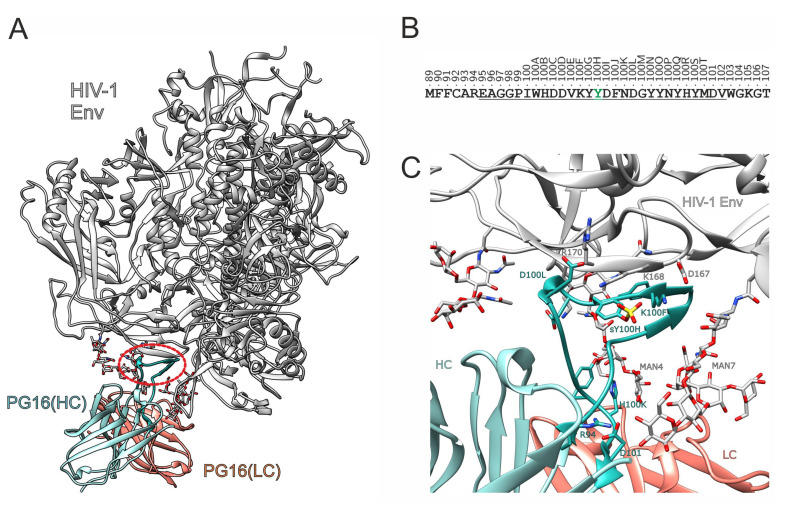
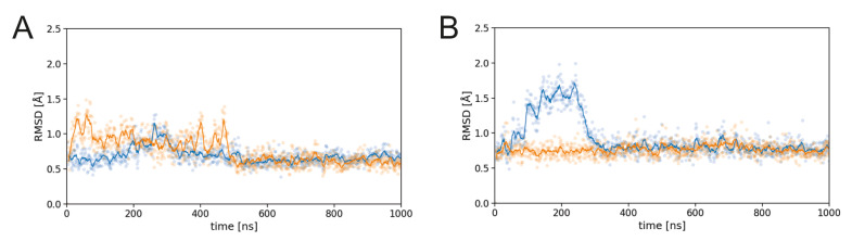
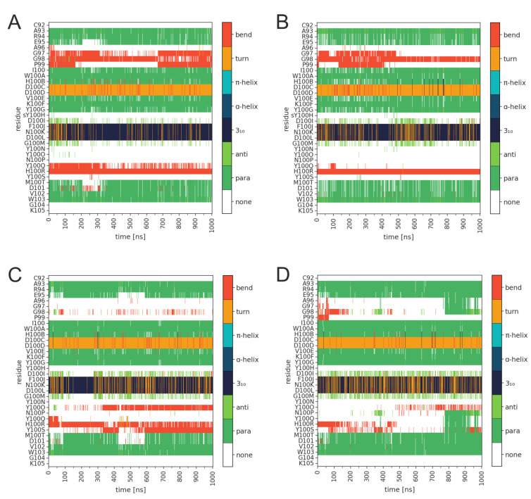
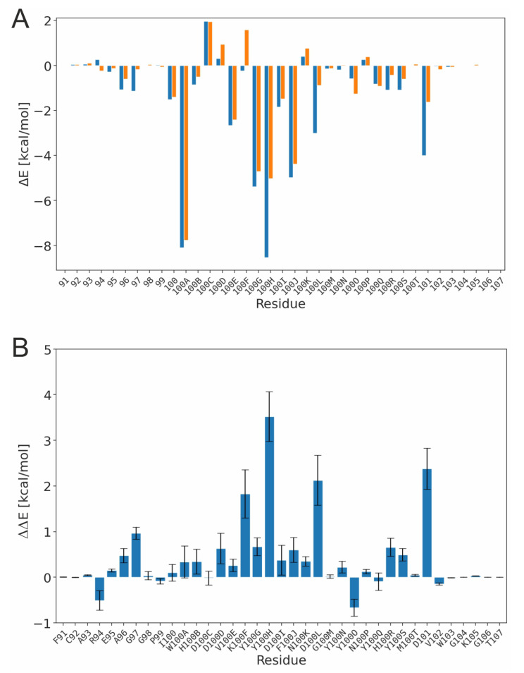
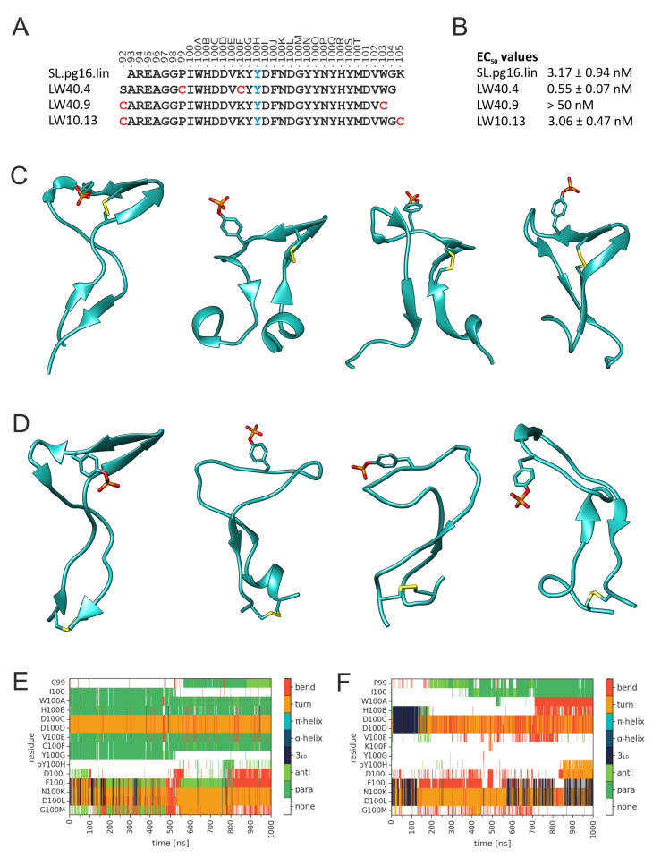
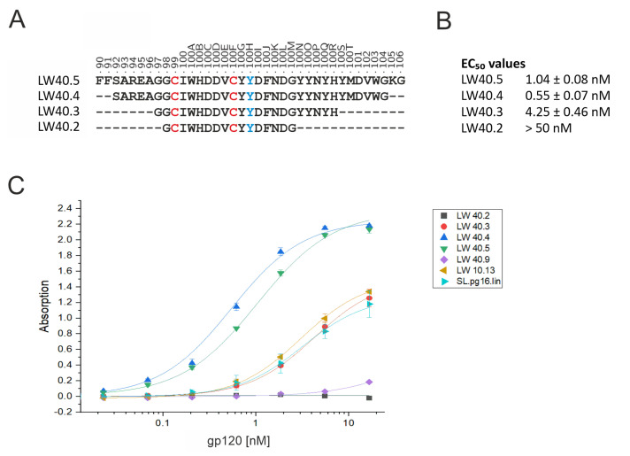
PG16 is a broadly neutralizing antibody that binds to the gp120 subunit of the HIV-1 Env protein. The major interaction site is formed by the unusually long complementarity determining region (CDR) H3. The CDRH3 residue Tyr100H is known to represent a tyrosine sulfation site; however, this modification is not present in the experimental complex structure of PG16 with full-length HIV-1 Env. To investigate the role of sulfation for this complex, we modeled the sulfation of Tyr100H and compared the dynamics and energetics of the modified and unmodified complex by molecular dynamics simulations at the atomic level. Our results show that sulfation does not affect the overall conformation of CDRH3, but still enhances gp120 interactions both at the site of modification and for the neighboring residues. This stabilization affects not only protein-protein contacts, but also the interactions between PG16 and the gp120 glycan shield. Furthermore, we also investigated whether PG16-CDRH3 is a suitable template for the development of peptide mimetics. For a peptide spanning residues 93-105 of PG16, we obtained an experimental EC50 value of 3nm for the binding of gp120 to the peptide. This affinity can be enhanced by almost one order of magnitude by artificial disulfide bonding between residues 99 and 100F. In contrast, any truncation results in significantly lower affinity, suggesting that the entire peptide segment is involved in gp120 recognition. Given their high affinity, it should be possible to further optimize the PG16-derived peptides as potential inhibitors of HIV invasion.

Keywords: HIV-1; PG16; antibody; antibody mimetic peptides; molecular dynamics; peptides.


Conflict of interest statement

The authors declare no conflict of interest.

Figures

Figure 1
Figure 1
Structure of the PG16-Env complex. (A) Experimental structure of the PG16-Env complex (PDB code: 6ULC; [16]). The heavy and light chain of the antibody are shown in cyan and orange, respectively, and HIV-1 Env is shown in white. The red oval frames the CDRH3, which adopts the form of a hammerhead. Glycans are shown in stick presentation. (B) Sequence of PG16-CDRH3 (underlined) including flanking residues. The positions are labeled using the Kabat numbering scheme for antibodies [17]. Position 100H highlighted in green marks the tyrosine residue that can be post-translationally sulfated. (C) Enlargement of the binding site showing the interactions of CDRH3. Important residues of CDRH3 are shown as cyan sticks; interacting gp120 residues and glycans are colored according to their atom types. The sulfo group is modeled in the structure to illustrate its putative position in the interface.
Figure 2
Figure 2
Dynamics of PG16-CDRH3 in the PG16-Env complex. RMSD for (A) TYS-PG16 and (B) TYR-PG16. Data for the first and second simulation run are shown in blue and orange, respectively. In both plots, explicit values are highlighted as dots and running averages are shown as lines.
Figure 3
Figure 3
Secondary structure of the PG16-CDRH3 in the PG16-Env complex for (A,B) TYS-PG16 and (C,D) TYR-PG16. The two panels for each system show the two independent simulation runs. The color code for the different types of secondary structure is given in the bar on the right.
Figure 4
Figure 4
Energetics of the PG16-CDRH3 in the PG16-Env complex. (A) Interaction energy derived from a per-residue binding energy decomposition. Values for TYS-PG16 and TYR-PG16 are shown in blue and orange, respectively. Negative values indicate a favorable contribution to binding. Values are averaged over two simulations runs for each system. (B) Differences in the energetic contributions of individual residues between TYS-PG16 and TYR-PG16. Positive values indicate a stronger interaction of the respective residue in TYS-PG16. The standard error is indicated by error bars.
Figure 5
Figure 5
Key interactions of PG16 residue 100H. (A) Intermolecular interaction of sY100H with K168 of gp120. (B) Shortest distance between the sY100H sulfo group and the K168 ammonium group (run1, blue; run2, orange). Explicit values are highlighted by dots and running averages are shown as lines. (C) Shortest distance between the Y100H sidechain hydroxyl group and the K168 ammonium group. (D) Intramolecular interaction of sY100H with K100F of PG16. (E) Shortest distance between the sY100H sulfo group and the K100F ammonium group (run1, blue; run2, orange). (F) Shortest distance between the Y100H sidechain hydroxyl group and the K100F ammonium group.
Figure 6
Figure 6
Key interactions of PG16 residues K100F and D100L. (A) Intermolecular interaction of K100F with D167 of gp120 (black dotted line). In addition, the intramolecular sY100H-K100F interaction is indicated by a white dotted line. (B,C) Shortest distance between the K100F ammonium group and the D167 carboxy group in (B) TYS-PG16 and (C) TYR-PG16 (run1, blue; run2, orange). Explicit values are highlighted as dots and running averages are shown as lines. (D) Interaction of D100L with R170 of gp120. (C,D) Shortest distance between the D100L carboxy group and the R170 guanidino group in (E) TYS-PG16 and (F) TYR-PG16 (run1, blue; run2, orange).
Figure 7
Figure 7
Key interactions of PG16 residues H100R and D101. (A) Interaction of H100R with the MAN4 glycan of gp120 (black dotted line). (B,C) Shortest distance between the H100R imidazole ring and the MAN4 glycan in (B) TYS-PG16 and (C) TYR-PG16 (run1, blue; run2, orange). Explicit values are highlighted as dots and running averages are shown as lines. (D) Interaction of D101 with the MAN7 glycan of gp120. (E,F) Shortest distance between the D100 carboxyl oxygens and the hydroxy groups of the MAN7 glycan (E) TYS-PG16 and (F) TYR-PG16 (run1, blue; run2, orange). The two shades of blue and orange indicate the interactions of the individual oxygens, Oδ1 and Oδ2. The black dotted line marks a distance of 2.5Å, as the upper limit for the formation of a hydrogen bond (measured between the oxygen atom as the donor and hydrogen atom as the hydrogen bond acceptor).
Figure 8
Figure 8
Effect of disulfide bonds on the stability of PG16-CDRH3-derived peptides. (A) Schematic presentation of the alternative disulfide bonding patterns investigated. Cysteines forming an intramolecular disulfide bond are highlighted in red. Position 100H highlighted in blue represents a phosphotyrosine. (B) Experimental EC50 values for binding of HIV-1 gp120 (HxBc2) to the disulfide-bonded peptides in comparison to the linear peptide SL.pg16.lin. (C) Snapshots from the MD simulation illustrating the dynamics of the free LW40.04 peptide. The initial structure is shown in the left panel. The position of the disulfide bond and of the phosphotyrosine is indicated as sticks. (D) Snapshots from the MD simulation illustrating the dynamics of the free LW40.9 peptide. The initial structure is shown in the left panel. The position of the disulfide bond and of the phosphotyrosine is indicated as sticks. (E,F) Conformational stability of the central β-hairpin in (E) LW40.4 and (F) LW40.9. The color code for the different types of secondary structure is given in the bar on the right.
Figure 9
Figure 9
Effect of peptide length and disulfide bridges on the binding properties of PG16−CDRH3−derived peptides. (A) Sequence of the four peptides LW40.5, LW40.4, LW40.3, and LW40.2, which differ in their length of the N- and C-termini. Cysteines forming an intramolecular disulfide bond are highlighted in red. Position 100H highlighted in blue represents a phosphotyrosine. (B) Experimental EC50 values for binding of HIV-1 gp120 (HxBc2) to the peptides of different length. (C) Dose-dependent binding of gp120 binding to various PG16-derived peptides (see Table 1 for a complete list of peptide sequences). See Materials and Methods for details (Section 2.4).

Similar articles

Cited by

References

    1. Barré-Sinoussi F., Chermann J.C., Rey F., Nugeyre M.T., Chamaret S., Gruest J., Dauguet C., Axler-Blin C., Vézinet-Brun F., Rouzioux C., et al. Isolation of a T-lymphotropic retrovirus from a patient at risk for acquired immune deficiency syndrome (AIDS) Science. 1983;220:868–871. doi: 10.1126/science.6189183. - DOI - PubMed
    1. Gallo R.C., Salahuddin S.Z., Popovic M., Shearer G.M., Kaplan M., Haynes B.F., Palker T.J., Redfield R., Oleske J., Safai B., et al. Frequent detection and isolation of cytopathic retroviruses (HTLV-III) from patients with AIDS and at risk for AIDS. Science. 1984;224:500–503. doi: 10.1126/science.6200936. - DOI - PubMed
    1. UNAIDS Homepage—unaids.org. 2023. [(accessed on 23 April 2023)]. Available online: https://www.unaids.org/en.
    1. Spencer D.A., Shapiro M.B., Haigwood N.L., Hessell A.J. Advancing HIV broadly neutralizing antibodies: From discovery to the clinic. Front. Public Health. 2021;9:690017. doi: 10.3389/fpubh.2021.690017. - DOI - PMC - PubMed
    1. Walsh S.R., Seaman M.S. Broadly neutralizing antibodies for HIV-1 prevention. Front. Immunol. 2021;12:712122. doi: 10.3389/fimmu.2021.712122. - DOI - PMC - PubMed

LinkOut - more resources