Review
. 2023 Jun 15;12(6):864.
doi: 10.3390/biology12060864.

The ACSL4 Network Regulates Cell Death and Autophagy in Diseases

Affiliations
Review

The ACSL4 Network Regulates Cell Death and Autophagy in Diseases

Fangquan Chen et al. Biology (Basel). .

Abstract

Lipid metabolism, cell death, and autophagy are interconnected processes in cells. Dysregulation of lipid metabolism can lead to cell death, such as via ferroptosis and apoptosis, while lipids also play a crucial role in the regulation of autophagosome formation. An increased autophagic response not only promotes cell survival but also causes cell death depending on the context, especially when selectively degrading antioxidant proteins or organelles that promote ferroptosis. ACSL4 is an enzyme that catalyzes the formation of long-chain acyl-CoA molecules, which are important intermediates in the biosynthesis of various types of lipids. ACSL4 is found in many tissues and is particularly abundant in the brain, liver, and adipose tissue. Dysregulation of ACSL4 is linked to a variety of diseases, including cancer, neurodegenerative disorders, cardiovascular disease, acute kidney injury, and metabolic disorders (such as obesity and non-alcoholic fatty liver disease). In this review, we introduce the structure, function, and regulation of ACSL4; discuss its role in apoptosis, ferroptosis, and autophagy; summarize its pathological function; and explore the potential implications of targeting ACSL4 in the treatment of various diseases.

Keywords: ACSL4; apoptosis; autophagy; cancer; ferroptosis.


Conflict of interest statement

The authors declare no conflict of interest.

Figures

Figure 1
Figure 1
Autophagy-dependent ferroptosis. Macroautophagy/autophagy or selective autophagy (lipophagy, ferritinophagy, clockophagy, chaperone-mediated autophagy) promotes the degradation of organelles, ferritin, lipid droplets, or proteins (such as GPX4 and ARNTL) to increase intracellular Fe2+ or free fatty acids, promoting ferroptosis. In addition, BECN1 binding to SLC7A11 or cathepsin B release also promotes ferroptosis through lipid peroxidation. In this process, ACSL4 plays a crucial role by effectively binding long-chain polyunsaturated fatty acids (PUFAs) with coenzyme A, enabling their re-esterification into phospholipids through the action of lysophosphatidylcholine acyltransferase 3 (LPCAT3). This interaction further facilitates the promotion of ferroptosis.
Figure 2
Figure 2
Structure of the ACSL4 gene and protein. (A) Simplified diagram of the ACSL4 gene. (B) Simplified diagram of the ACSL4 protein (top) and 3D model of ACSL4 protein.
Figure 3
Figure 3
Function of ACSL4. ACSL4 catalyzes the conversion of exogenous long-chain fatty acids into acyl-coenzyme A. Acyl-CoA can then be transported into the mitochondria via carnitine palmitoyltransferase-1 (CPT1), where it can be converted to acetyl-CoA and participate in the tricarboxylic acid cycle to provide energy. Additionally, acyl-CoA can produce phosphatidylinositol, which contributes to membrane synthesis. Acyl-CoA can promote diacylglycerol acyltransferase (DGAT) and be involved in the synthesis of triacylglycerol (TAG) in lipid droplets (LDs) for lipid storage. Intermediates such as lyso-phosphatidic acid (LPA), phosphatidic acid (PA), and diacylglycerol (DAG) may initiate signaling cascades, and PA and DAG can also serve as precursors to all glycerophospholipids.
Figure 4
Figure 4
Transcriptional regulation of ACSL4. ACSL4 expression can be regulated by multiple transcription factors and non-coding RNA molecules (such as lncRNA and miRNA). Additionally, ACSL4 can undergo various post-translational modifications, including ubiquitination (Ub), phosphorylation (P), acetylation (ac), and O-GlcNAcylation (O-Glc), as well as engage in protein–protein interactions. These regulatory mechanisms play a crucial role in modulating cell death and various biological functions.
Figure 5
Figure 5
ACSL4 in apoptosis. On one hand, ACSL4 promotes apoptosis resistance by synthesizing lipids to increase mitochondrial membrane stability and by upregulating fatty-acid β-oxidation through ACSL4 expression. On the other hand, ACSL4 can also catalyze the synthesis of fatty acyl-CoA, which can generate lipid-free radicals and cause oxidative stress.
Figure 6
Figure 6
ACSL4 in ferroptosis. Exogenous and endogenous fatty acids undergo lipid peroxidation through ACSL4 to promote ferroptosis. The expression of ACSL4 is regulated by various pathways, including non-coding RNA, PKCβII, MAPK, and E-cadherin. Additionally, IFNγ and arachidonic acid from T cells in the tumor microenvironment promote ACSL4-dependent ferroptosis. On the other hand, there are multiple antioxidant pathways to inhibit ferroptosis. ACL: ATP-citrate lyase; POR, cytochrome p450 oxidoreductase; FASN, fatty acid synthase; GSH, glutathione; GCH1, GTP cyclohydrolase 1; BH4, tetrahydrobiopterin; AIFM2, apoptosis-inducing factor mitochondrial 2; CoQH2, ubiquinol; KEAP1, kelch-like ECH-associated protein 1; NFE2L2, nuclear factor erythroid 2-related factor 2; DHODH, dihydroorotate dehydrogenase.
Figure 7
Figure 7
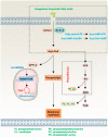
ACSL4 in autophagy. ACSL4 can regulate the autophagy process through different mechanisms. It can inhibit autophagy by promoting mTOR expression or binding to V-ATPase. On the other hand, Faa1, located in the phagophore, promotes de novo synthesis of fatty acids, and extracellular SQSTM1-dependent ACSL4 expression promotes autophagy.
Figure 8
Figure 8
The expression of ACSL4 in different tumors. *: p < 0.05; ***: p < 0.001. Data from http://timer.cistrome.org/ (accessed on 29 March 2023).

Similar articles

Cited by

References

    1. Overmyer K., Brosché M., Kangasjärvi J. Reactive oxygen species and hormonal control of cell death. Trends Plant Sci. 2003;8:335–342. doi: 10.1016/S1360-1385(03)00135-3. - DOI - PubMed
    1. Livesey K.M., Kang R., Vernon P., Buchser W., Loughran P., Watkins S.C., Zhang L., Manfredi J.J., Zeh H.J., 3rd, Li L., et al. p53/HMGB1 complexes regulate autophagy and apoptosis. Cancer Res. 2012;72:1996–2005. doi: 10.1158/0008-5472.CAN-11-2291. - DOI - PMC - PubMed
    1. Yin X., Cao L., Kang R., Yang M., Wang Z., Peng Y., Tan Y., Liu L., Xie M., Zhao Y., et al. UV irradiation resistance-associated gene suppresses apoptosis by interfering with BAX activation. EMBO Rep. 2011;12:727–734. doi: 10.1038/embor.2011.79. - DOI - PMC - PubMed
    1. Dixon S.J., Lemberg K.M., Lamprecht M.R., Skouta R., Zaitsev E.M., Gleason C.E., Patel D.N., Bauer A.J., Cantley A.M., Yang W.S., et al. Ferroptosis: An iron-dependent form of nonapoptotic cell death. Cell. 2012;149:1060–1072. doi: 10.1016/j.cell.2012.03.042. - DOI - PMC - PubMed
    1. Chen X., Kang R., Kroemer G., Tang D. Broadening horizons: The role of ferroptosis in cancer. Nat. Rev. Clin. Oncol. 2021;18:280–296. doi: 10.1038/s41571-020-00462-0. - DOI - PubMed

LinkOut - more resources