. 2023 Jun 16;12(6):873.
doi: 10.3390/biology12060873.

Derivatives of Differentiation-Inducing Factor 1 Differentially Control Chemotaxis and Stalk Cell Differentiation in Dictyostelium discoideum

Affiliations

Derivatives of Differentiation-Inducing Factor 1 Differentially Control Chemotaxis and Stalk Cell Differentiation in Dictyostelium discoideum

Hidekazu Kuwayama et al. Biology (Basel). .

Abstract

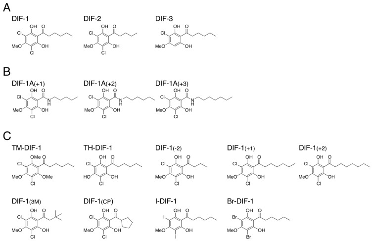
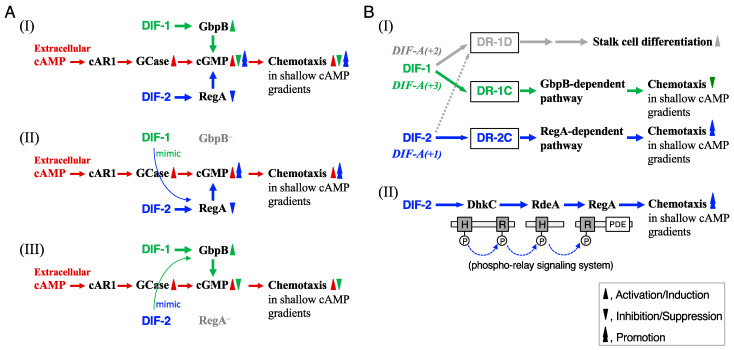
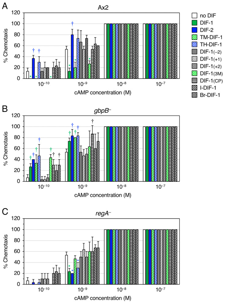
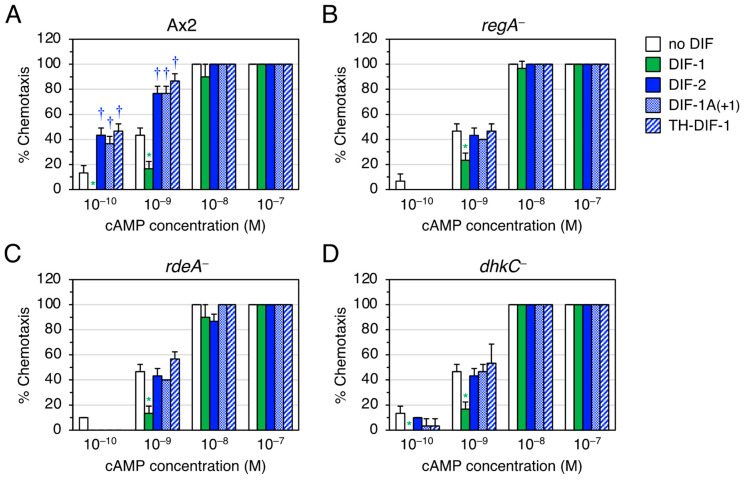
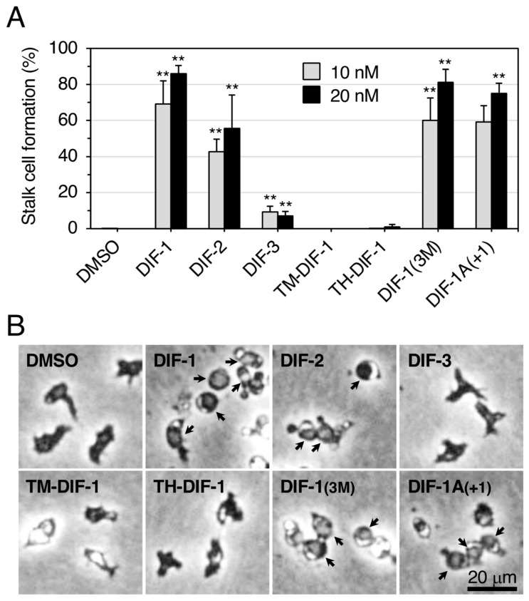
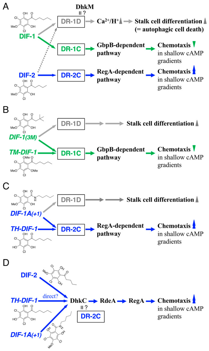
Differentiation-inducing factors 1 and 2 (DIF-1 and DIF-2) are small lipophilic signal molecules that induce stalk cell differentiation but differentially modulate chemotaxis toward cAMP in the cellular slime mold Dictyostelium discoideum; DIF-1 suppresses chemotactic cell movement in shallow cAMP gradients, whereas DIF-2 promotes it. The receptor(s) for DIF-1 and DIF-2 have not yet been identified. We examined the effects of nine derivatives of DIF-1 on chemotactic cell movement toward cAMP and compared their chemotaxis-modulating activity and stalk cell differentiation-inducing activity in wild-type and mutant strains. The DIF derivatives differentially affected chemotaxis and stalk cell differentiation; for example, TM-DIF-1 suppressed chemotaxis and showed poor stalk-inducing activity, DIF-1(3M) suppressed chemotaxis and showed strong stalk-inducing activity, and TH-DIF-1 promoted chemotaxis. These results suggest that DIF-1 and DIF-2 have at least three receptors: one for stalk cell induction and two for chemotaxis modulation. In addition, our results show that the DIF derivatives can be used to analyze the DIF-signaling pathways in D. discoideum.

Keywords: DIF; DhkC; Dictyostelium; GbpB; RdeA; RegA; chemotaxis; stalk cell differentiation.


Conflict of interest statement

The authors declare no conflicts of interest.

Figures

Figure 1
Figure 1
Chemical structure of (A) DIFs 1–3, (B) amide derivatives of DIF-1, and (C) the nine DIF derivatives used in this study. Note that DIF-1A(+1), DIF-1A(+2), and DIF-1A(+3) were referred to as DIF-1[A+1], DIF-1[A+2], and DIF-1[A+3] under our previous nomenclature [11].
Figure 2
Figure 2
(A) Proposed scheme for the actions of DIF-1 and DIF-2 in modulating chemotaxis [10]. (I) Dictyostelium cells show chemotactic movement toward extracellular cAMP, which induces chemotaxis by binding to the cell surface cAMP receptor (cAR1), followed by activation of guanylyl cyclase (GCase) and an increase in intracellular cGMP. In shallow cAMP gradients, DIF-1 inhibits chemotaxis toward cAMP, at least in part, via activation of the cGMP-PDE GbpB and a subsequent decrease in intracellular cGMP, whereas DIF-2 enhances chemotaxis, at least in part, via a RegA (a cAMP-PDE)-dependent pathway and a subsequent increase in intracellular cGMP. However, at high concentrations of DIFs (e.g., 100 nM), cross-talk can occur, and DIF-1 and DIF-2 both enhance chemotaxis in gbpB cells (II) and inhibit chemotaxis in regA cells (III). (B) (I) Proposed scheme for the actions of DIF compounds in inducing stalk cell differentiation and modulating chemotaxis via three putative DIF receptors [11,20,21]. During normal development, DIF-1 would induce stalk cell differentiation, at least in part, via a DIF receptor (DR-1D) and negatively modulate chemotaxis via another DIF receptor (DR-1C) and a GbpB-dependent pathway. In contrast, DIF-2 would function mainly as a positive modulator for chemotaxis, at least in part, via another DIF receptor (DR-2C) and a RegA-dependent pathway. The artificial compounds, DIF-1A(+2) and DIF-1A(+3), would be efficient stalk cell inducers and chemotaxis modulators, possibly via DR-1D and DR-1C, respectively. DIF-1A(+1), like DIF-2, would induce stalk cell differentiation via DR-1D and modulate chemotaxis via DR-2C. Note that the DIF receptors that were DR-1, DR-2 and DR-3 under our previous nomenclature [11] are referred to as DR-1D, DR-1C, and DR-2C, respectively, in our previous [21] and present study in order to match the names of the receptors and their ligands, DIF-1 and DIF-2. (II) Proposed scheme for the actions of DIF-2 via the DhkC–RdeA–RegA phospho-relay pathway. The schematic diagram of the phospho-relay pathway illustrates the previously proposed model [22,23,24,25]; DhkC, Dictyostelium histidine kinase C, phosphorylates itself and passes the phosphate through the relay by RdeA to RegA, resulting in activation of RegA (cAMP phosphodiesterase). DIF-2 modulates chemotaxis, at least in part, via the Dictyostelium phospho-relay signaling system, DhkC–RdeA–RegA pathway [20]. H, a site of histidine phosphorylation. R, receiver domain. PDE, phosphodiesterase. The catalytic domain of DhkC is omitted for simplicity.
Figure 3
Figure 3
Effect of DIFs (100 nM) on chemotaxis in Ax2, gbpB, and regA cells. (A) Ax2, (B) gbpB, and (C) regA cells were starved for 6 h in shake-culture, and cell droplets were spotted on PB agar containing 3 mM caffeine (Control) plus 100 nM DIF compounds. Cells were assayed for chemotaxis toward the indicated doses of cAMP; 10 cell droplets were examined for each cAMP concentration. Data are presented as the mean ± SD of triplicate measurements (n = 3) for one experiment. * and † signify statistically significant inhibition and promotion of chemotaxis, respectively; p < 0.05 versus Control.
Figure 4
Figure 4
Effect of DIFs (10 nM) on chemotaxis in Ax2, regA, rdeA, and dhkC cells. (A) Ax2, (B) regA, (C) rdeA, and (D) dhkC cells were starved for 6 h in shake-culture, and cell droplets were spotted on PB agar containing 3 mM caffeine (Control) plus 10 nM DIF compounds. Cells were assayed for chemotaxis toward the indicated doses of cAMP; 10 cell droplets were examined for each cAMP concentration. Data are presented as the mean ± SD of triplicate measurements (n = 3) for one experiment. * and † signify statistically significant inhibition and promotion of chemotaxis, respectively; p < 0.05 versus Control.
Figure 5
Figure 5
Effect of DIFs on stalk cell differentiation in HM1030 cells. (A) Cells were incubated without additives for 8 h, with ~4.2 mM cAMP for 16 h, and then with 0.1% or 0.2% DMSO (vehicle), or 10 nM or 20 nM DIF compounds for 24 h (total 48 h), and the stalk cells (% of total cells) were counted by using phase-contrast microscopy. Data are presented as the mean ± SD (bars) of three independent experiments (n = 3). ** p < 0.01 versus DMSO control. (B) Representative photos of cells after treatment with 0.1% DMSO or the indicated DIF compounds at 10 nM. Arrows indicate stalk cells.
Figure 6
Figure 6
(A) Chemical structures of DIF-1 and DIF-2 and scheme for their actions in inducing stalk cell differentiation and modulating chemotaxis. We assume here that DIF-1 would induce stalk cell differentiation via its receptor DR-1D and subsequent increases in cytoplasmic Ca2+ and H+ concentrations, at least in part [12,13,14,32], and that DIF-1 would negatively modulate chemotaxis in shallow cAMP gradients via another receptor, DR-1C, whereas DIF-2 would positively modulate chemotaxis in shallow cAMP gradients via its receptor DR-2C. Note that DhkM, another receptor-type Dictyostelium histidine kinase, is involved in DIF-1-induced stalk cell differentiation (autophagic cell death) [36], and DhkM might be DR-1D [37]. (B) Chemical structures of the DIF-1-type molecules, DIF-1(3M) and TM-DIF-1, and scheme for their actions in inducing stalk cell differentiation and inhibiting chemotaxis via the putative DIF receptors. (C) Chemical structures of the DIF-2-type molecules, DIF-1A(+1) and TH-DIF-1, and scheme for their actions in inducing stalk cell differentiation and promoting chemotaxis via the putative DIF receptors. (D) Chemical structures of DIF-2, DIF-1A(+1), and TH-DIF-1, and scheme for their actions in promoting chemotaxis via the DhkC–RdeA–RegA pathway. DhkC might be DR-2C [20].

References

    1. Konijn T.M., van de Meene J.G.C., Bonner J.T., Barkley D.S. The acrasin activity of adenosine-3′,5′-cyclic phosphate. Proc. Natl. Acad. Sci. USA. 1967;58:1152–1154. doi: 10.1073/pnas.58.3.1152. - DOI - PMC - PubMed
    1. Darmon M., Brachet P., Pereira da Silva L.H. Chemotactic signals induce cell differentiation in Dictyostelium discoideum. Proc. Natl. Acad. Sci. USA. 1975;72:3163–3166. doi: 10.1073/pnas.72.8.3163. - DOI - PMC - PubMed
    1. Devreotes P. Dictyostelium discoideum: A model system for cell-cell interactions in development. Science. 1989;245:1054–1058. doi: 10.1126/science.2672337. - DOI - PubMed
    1. Morris H.R., Taylor G.W., Masento M.S., Jermyn K.A., Kay R.R. Chemical structure of the morphogen differentiation inducing factor from Dictyostelium discoideum. Nature. 1987;328:811–814. doi: 10.1038/328811a0. - DOI - PubMed
    1. Morris H.R., Masento M.S., Taylor G.W., Jermyn K.A., Kay R.R. Structure elucidation of two differentiation inducing factors (DIF-2 and DIF-3) from the cellular slime mould Dictyostelium discoideum. Biochem. J. 1988;249:903–906. doi: 10.1042/bj2490903. - DOI - PMC - PubMed

LinkOut - more resources