Review
. 2023 Jun 17;12(6):878.
doi: 10.3390/biology12060878.

Emerging Rhabdoviruses and Human Infection

Affiliations
Review

Emerging Rhabdoviruses and Human Infection

James G Shepherd et al. Biology (Basel). .

Abstract

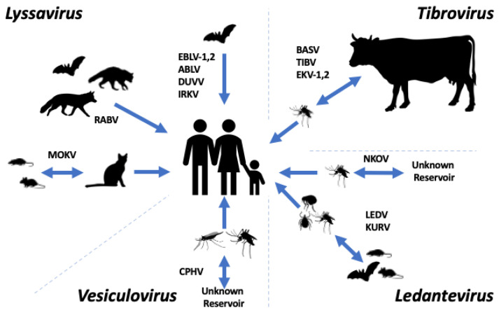
Rhabdoviridae is a large viral family, with members infecting a diverse range of hosts including, vertebrate species, arthropods, and plants. The predominant human pathogen within the family is Rabies lyssavirus, the main cause of human rabies. While rabies is itself a neglected disease, there are other, less well studied, rhabdoviruses known to cause human infection. The increasing application of next-generation sequencing technology to clinical samples has led to the detection of several novel or rarely detected rhabdoviruses associated with febrile illness. Many of these viruses have been detected in low- and middle-income countries where the extent of human infection and the burden of disease remain largely unquantified. This review describes the rhabdoviruses other than Rabies lyssavirus that have been associated with human infection. The discovery of the Bas Congo virus and Ekpoma virus is discussed, as is the re-emergence of species such as Le Dantec virus, which has recently been detected in Africa 40 years after its initial isolation. Chandipura virus and the lyssaviruses that are known to cause human rabies are also described. Given their association with human disease, the viruses described in this review should be prioritised for further study.

Keywords: Rhabdoviridae; human infection; lyssavirus; rabies; viral zoonoses.


Conflict of interest statement

The authors declare no conflict of interest.

Figures

Figure 1
Figure 1
Transmission routes of selected members of the subfamily Alpharhabdovirinae. Depiction of the known and possible transmission routes for the members of the Lyssavirus genus (Rabies virus (RABV), European bat lyssavirus-1 (EBLV-1), European bat lyssavirus-2 (EBLV-2), Australian bat lyssavirus (ABLV), Duvenhage virus (DUVV), Irkut virus (IRKV), and Mokola virus (MOKV)), the Tibrovirus genus (Bas-Congo virus (BASV), Tibrogargan virus (TIBV), Ekpoma virus-1 (EKV-1) and Ekpoma virus-2 (EKV-2)), Ledantevirus genus (Le Dantec virus (LEDV), Nkolbisson virus (NKOV), and Kumasi virus (KURV), and the Vesiculovirus genus (Chandipura vesiculovirus (CHPV)).
Figure 2
Figure 2
Rhabdovirus structure and genome organisation. (A) Structure of the prototype rhabdovirus Vesicular Stomatitis Virus (VSV). (B) Genome organisation of the canonical rhabdovirus VSV (accession number J02428) demonstrating the canonical genes in 3′-5′ direction: nucleoprotein (N), phosphoprotein (P), matrix protein (M), glycoprotein (G), and the large protein comprising the RDRP (L).
Figure 3
Figure 3
The sub-family Alpharhabdovirinae. Maximum likelihood phylogeny of the sub-family Alpharhabdovirinae based on a trimmed alignment of full-length L protein amino acid sequences. Viruses associated with human infection are indicated in orange text. The scale bar represents amino acid substitutions per site. Black circles indicate nodes with support values greater than 70 based on 1000 bootstrap replicates.

References

    1. Amarasinghe G.K., Ayllón M.A., Bào Y., Basler C.F., Bavari S., Blasdell K.R., Briese T., Brown P.A., Bukreyev A., Balkema-Buschmann A., et al. Taxonomy of the order Mononegavirales: Update 2019. Arch. Virol. 2019;164:1967–1980. doi: 10.1007/s00705-019-04247-4. - DOI - PMC - PubMed
    1. Dietzgen R.G., Kondo H., Goodin M.M., Kurath G., Vasilakis N. The family Rhabdoviridae: Mono—And bipartite negative-sense RNA viruses with diverse genome organization and common evolutionary origins. Virus Res. 2017;227:158–170. doi: 10.1016/j.virusres.2016.10.010. - DOI - PMC - PubMed
    1. Ge P., Tsao J., Schein S., Green T.J., Luo M., Zhou Z.H. Cryo-EM model of the bullet-shaped vesicular stomatitis virus. Science. 2010;327:689–693. doi: 10.1126/science.1181766. - DOI - PMC - PubMed
    1. Roche S., Bressanelli S., Rey F.A., Gaudin Y. Crystal Structure of the Low-pH Form of the Vesicular Stomatitis Virus Glycoprotein G. Science. 2006;313:187–191. doi: 10.1126/science.1127683. - DOI - PubMed
    1. Roche S., Rey F.A., Gaudin Y., Bressanelli S. Structure of the Prefusion Form of the Vesicular Stomatitis Virus Glycoprotein G. Science. 2007;315:843–848. doi: 10.1126/science.1135710. - DOI - PubMed

LinkOut - more resources