Case Reports
. 2023 May 29;14(6):1193.
doi: 10.3390/genes14061193.

A Carrier Female Manifesting an Unusual X-Linked Retinoschisis Phenotype Associated with the Pathogenic Variant c.266delA, p.(Tyr89LeufsTer37) in RS1, and Skewed X-Inactivation

Affiliations
Case Reports

A Carrier Female Manifesting an Unusual X-Linked Retinoschisis Phenotype Associated with the Pathogenic Variant c.266delA, p.(Tyr89LeufsTer37) in RS1, and Skewed X-Inactivation

Jennifer Kirkby et al. Genes (Basel). .

Abstract

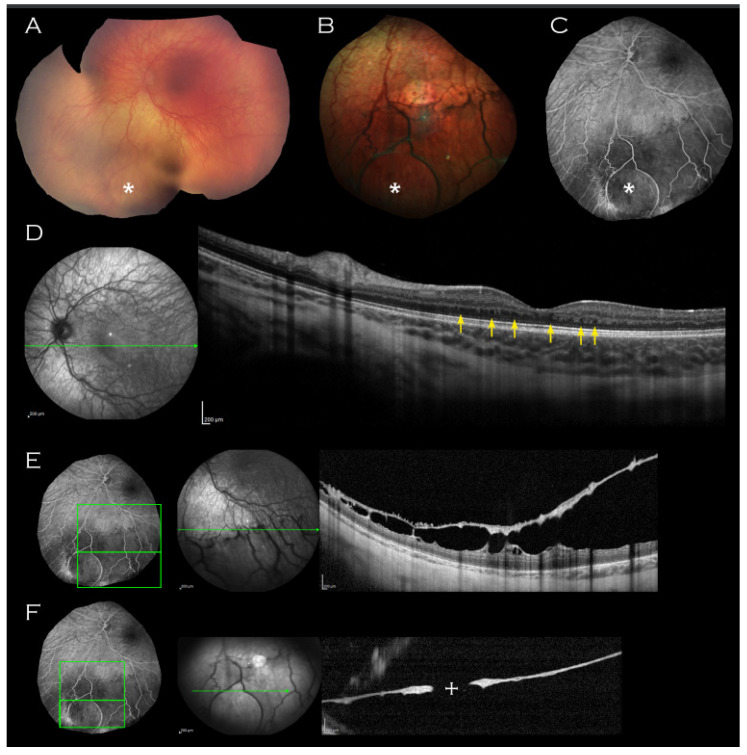
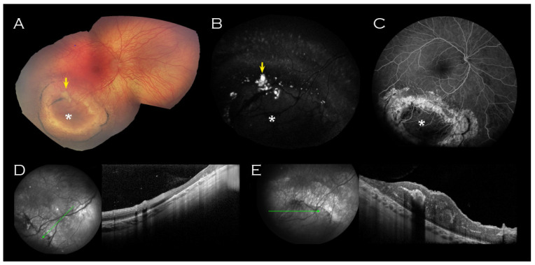
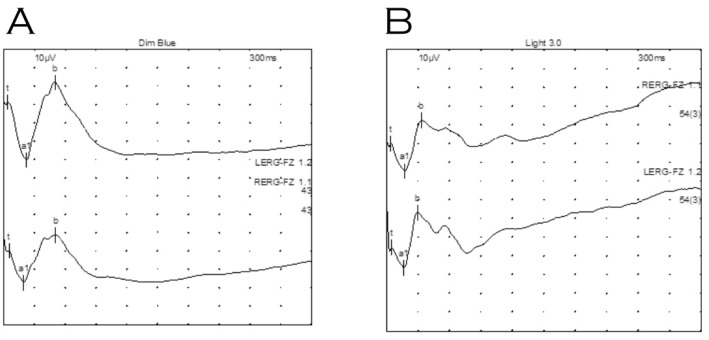
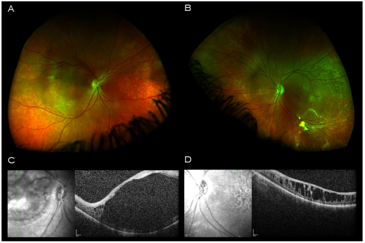
X-linked retinoschisis (XLRS) is the most common juvenile macular degeneration in males. Unlike most other X-linked retinal dystrophies, carrier heterozygous females are very rarely reported to show clinical features of the disease. Herein, we describe unusual retinal features in a 2-year-old female infant with family history and genetic testing consistent with XLRS.

Keywords: X-inactivation; X-linked retinoschisis; carrier; female; heterozygous.


Conflict of interest statement

The authors declare no conflict of interest. The funders had no role in the design of the study; in the collection, analyses, or interpretation of data; in the writing of the manuscript, or in the decision to publish the results.

Figures

Figure 1
Figure 1
Left eye imaging of proband. (A) Retcam montage, (B) colour fundus photograph and (C) fluorescein angiogram shows circular area of thinned elevated retina (indicated by asterisk) seen consistently on all imaging modalities. (D) Macular OCT demonstrating unusual thickening of the retina with absence of laminal structure and minute intraretinal hyporeflective cystic lesions (indicated by arrows) and (E,F) OCT through the lesion with approximate mapping of location on fluorescein angiogram image. An associated outer leaf break is demonstrated ((F), cross).
Figure 2
Figure 2
Right eye imaging of proband. Inferotemporal peripheral island of abnormal retina (indicated by asterisk) on (A) Retcam montage, (B) fundus autofluorescence and (C) fluorescein angiogram. Associated yellow material (arrow (A,B)) was hyperfluorescent on fundus autofluorescence. (D) OCT of the surrounded atrophic retina corresponded with thinned retina and (E) OCT through the centre of the lesion showed thickened retina corresponding to the relatively hypofluorescent island seen on FFA. No schisis was demonstrated on OCT through the lesion.
Figure 3
Figure 3
Electroretinography of proband under general anaesthetic. (A) Dark adapted dim blue scotopic response. (B) Light adapted standard flash (LA 3.0). Waveforms were not electronegative, however, waveforms for right eye (top) indicate relatively reduced b wave amplitudes compared to those for the left eye (bottom).
Figure 4
Figure 4
Imaging of proband’s father. Wide field retinal imaging (A) right eye (B) and left eye shows mild retinal pigment epithelium changes at the maculae and left vitreous veil at left inferior arcade. (C) Macular OCT right eye demonstrating a central hyporeflective intraretinal cavity with adjacent small cystic-like hyporeflective intraretinal lesions peripherally. (D) Macular OCT left eye demonstrating large cystic-like hyporeflective intraretinal lesions throughout, maximal in size and coalescing centrally.

References

    1. Sauer C.G., Gehrig A., Warneke-Wittstock R., Marquardt A., Ewing C.C., Gibson A., Lorenz B., Jurklies B., Weber B.H. Positional cloning of the gene associated with X-linked juvenile retinoschisis. Nat. Genet. 1997;17:164–170. doi: 10.1038/ng1097-164. - DOI - PubMed
    1. Molday R.S., Kellner U., Weber B.H. X-linked juvenile retinoschisis: Clinical diagnosis, genetic analysis, and molecular mechanisms. Prog. Retin. Eye Res. 2012;31:195–212. doi: 10.1016/j.preteyeres.2011.12.002. - DOI - PMC - PubMed
    1. Forsius H., Krause U., Helve J., Vuopala V., Mustonen E., Vainio-Mattila B., Fellman J., Eriksson A.W. Visual acuity in 183 cases of X-chromosomal retinoschisis. Can. J. Ophthalmol. 1973;8:385–393. - PubMed
    1. Sikkink S., Biswas S., Parry N.R.A., Stanga P.E., Trump D. X-linked retinoschisis: An update. J. Med. Genet. 2007;44:225–232. doi: 10.1136/jmg.2006.047340. - DOI - PMC - PubMed
    1. George N.D.L., Yates J.R.W., Moore A.T. Clinical Features in Affected Males With X-Linked Retinoschisis. Arch. Ophthalmol. 1996;114:274–280. doi: 10.1001/archopht.1996.01100130270007. - DOI - PubMed

Publication types