. 2023 Jun 11;24(12):10007.
doi: 10.3390/ijms241210007.

Dysregulation of Immune Cell Subpopulations in Atypical Hemolytic Uremic Syndrome

Affiliations

Dysregulation of Immune Cell Subpopulations in Atypical Hemolytic Uremic Syndrome

I-Ru Chen et al. Int J Mol Sci. .

Abstract

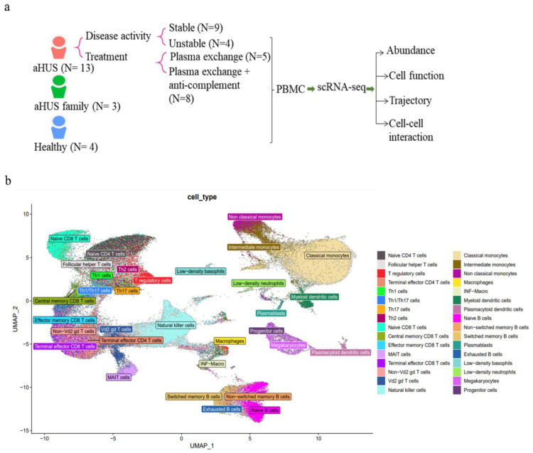
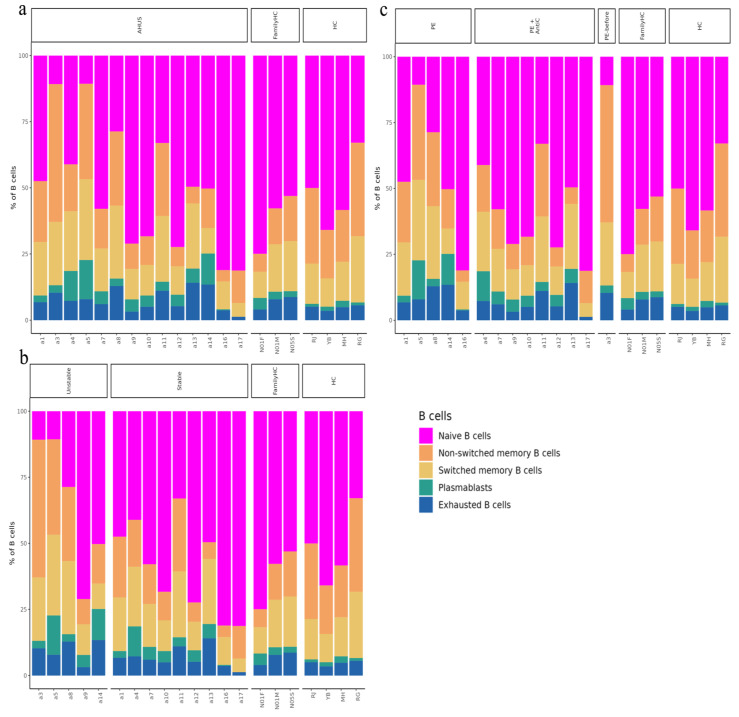
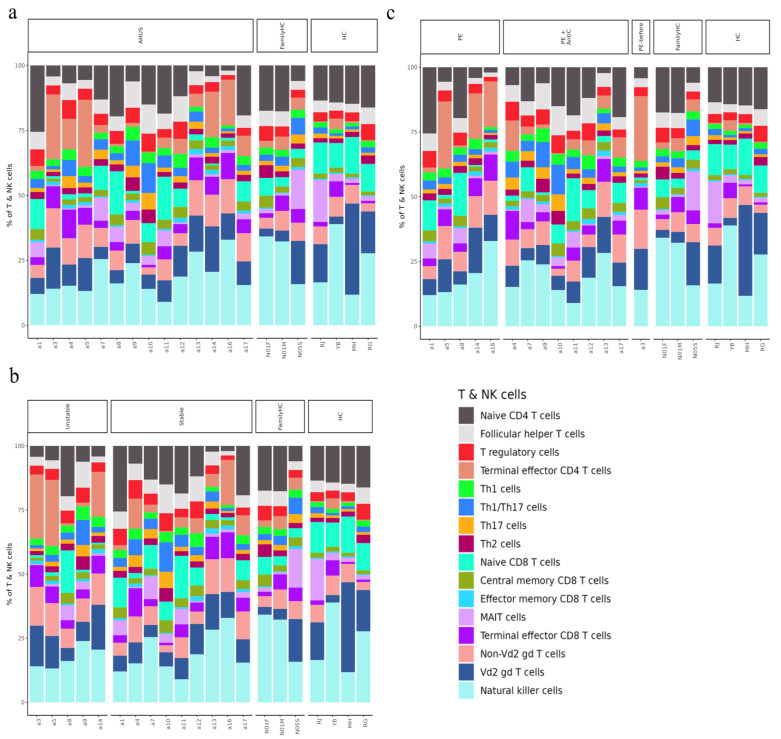
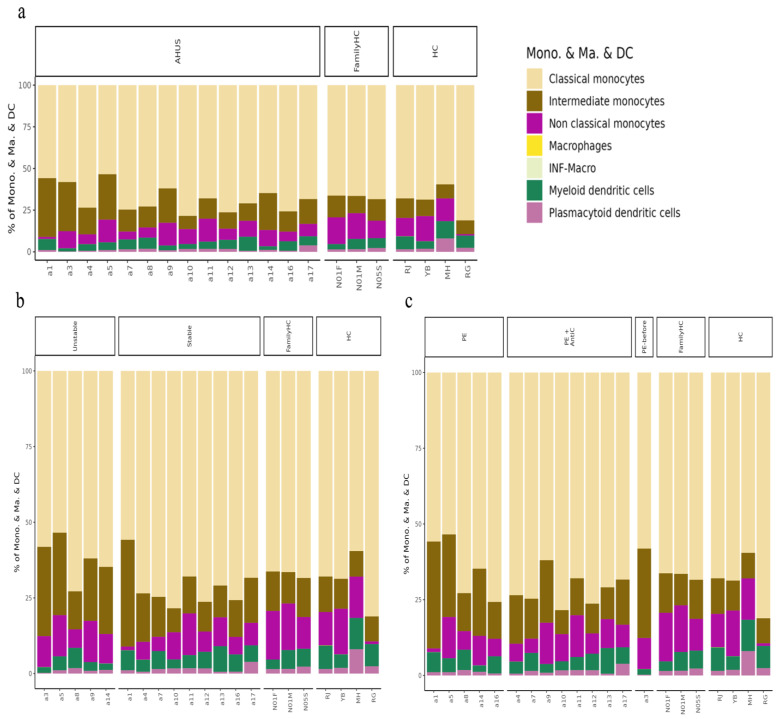
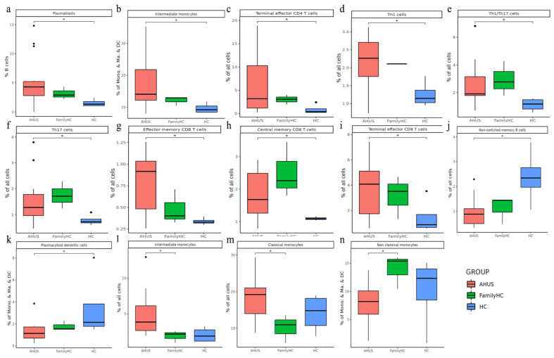
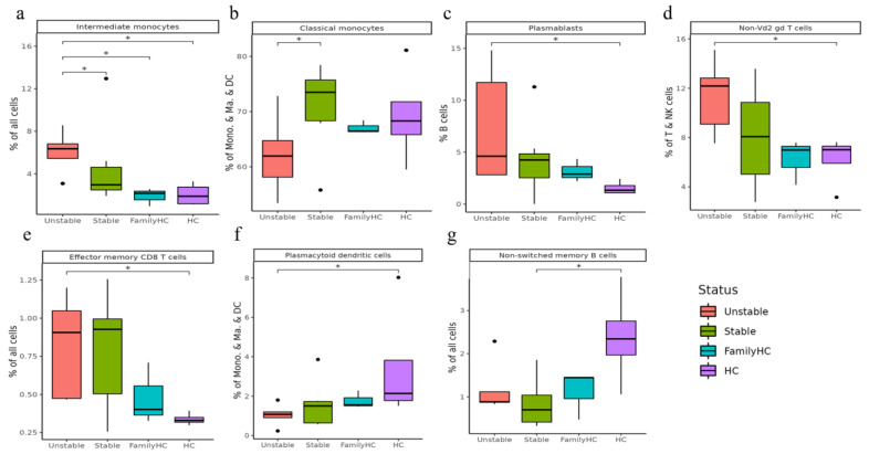
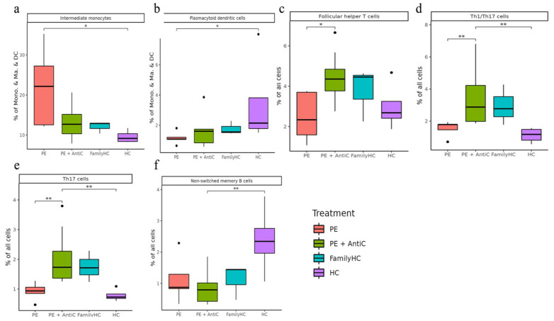
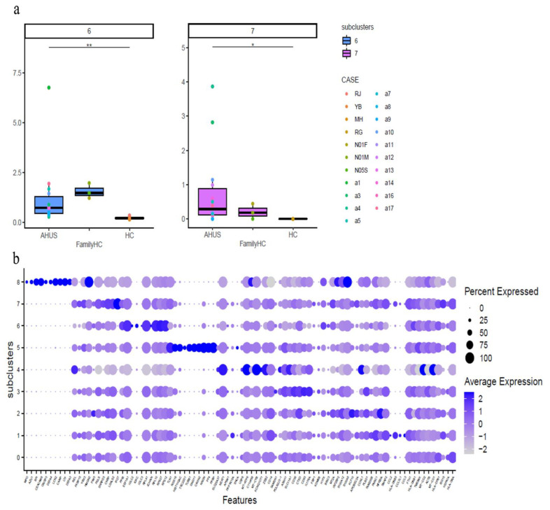
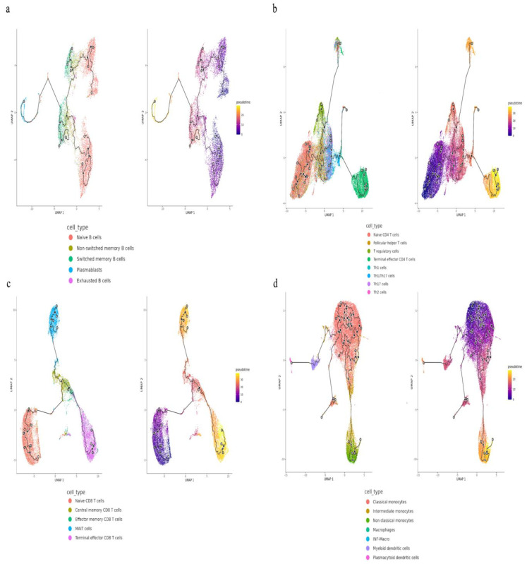
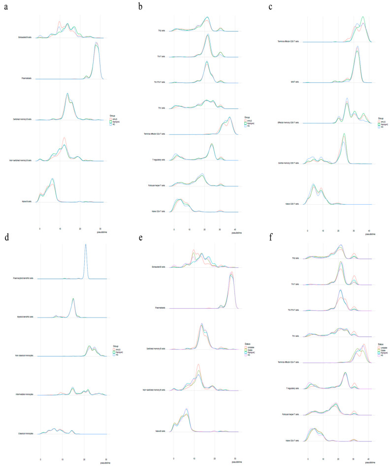
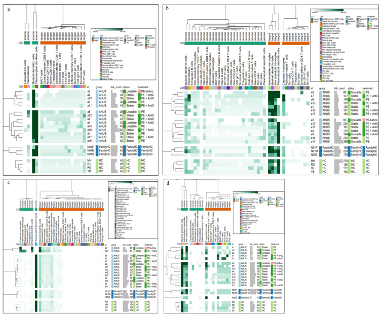
Atypical hemolytic uremic syndrome (aHUS) is a rare, life-threatening thrombotic microangiopathy. Definitive biomarkers for disease diagnosis and activity remain elusive, making the exploration of molecular markers paramount. We conducted single-cell sequencing on peripheral blood mononuclear cells from 13 aHUS patients, 3 unaffected family members of aHUS patients, and 4 healthy controls. We identified 32 distinct subpopulations encompassing 5 B-cell types, 16 T- and natural killer (NK) cell types, 7 monocyte types, and 4 other cell types. Notably, we observed a significant increase in intermediate monocytes in unstable aHUS patients. Subclustering analysis revealed seven elevated expression genes, including NEAT1, MT-ATP6, MT-CYB, VIM, ACTG1, RPL13, and KLRB1, in unstable aHUS patients, and four heightened expression genes, including RPS27, RPS4X, RPL23, and GZMH genes, in stable aHUS patients. Additionally, an increase in the expression of mitochondria-related genes suggested a potential influence of cell metabolism on the clinical progression of the disease. Pseudotime trajectory analysis revealed a unique immune cell differentiation pattern, while cell-cell interaction profiling highlighted distinctive signaling pathways among patients, family members, and controls. This single-cell sequencing study is the first to confirm immune cell dysregulation in aHUS pathogenesis, offering valuable insights into molecular mechanisms and potential new diagnostic and disease activity markers.

Keywords: atypical hemolytic uremic syndrome; complement; disease activity; single cell sequencing; therapy.


Conflict of interest statement

The authors declare no conflict of interest.

Figures

Figure 1
Figure 1
The immune cell phenotype of aHUS patients, aHUS family, and healthy controls were investigated using scRNA-seq analysis of PBMCs. The study design is presented in (a). UMAP coordinates showing the distribution of immune cells in PBMCs are presented in (b).
Figure 2
Figure 2
The relative abundance of subpopulations of B-cells is shown in (a), while the impact of aHUS disease activity on B-cell subpopulations is presented in (b), and the influence of aHUS treatment on B-cell subpopulations is shown in (c). The identifiers N01F, N01M, and N05S correspond to the individual cases of three aHUS family members included in our study. Similarly, the identifiers RJ, YB, MH, and RG denote the individual cases of four healthy controls in our study.
Figure 3
Figure 3
The relative abundance of subpopulations of T- and natural killer (NK) cells is shown in (a), the impact of aHUS disease activity on T- and NK cell subpopulations is presented in (b), and the influence of aHUS treatment on T- and NK cell subpopulations is shown in (c).
Figure 4
Figure 4
The analysis of monocyte series, including monocytes (Mono.), macrophages (Ma.), and dendritic cells (DC), is presented in (a), while the impact of aHUS disease activity on these cells is presented in (b), and the influence of aHUS treatment on these cells is shown in (c).
Figure 5
Figure 5
Significant abundance of cell subpopulations in PBMCs from aHUS, aHUS family, to healthy subjects (an) * p < 0.05.
Figure 6
Figure 6
Significant abundance of cell subpopulations in PBMCs from aHUS in unstable and stable disease activity, aHUS family, to healthy subjects (ag). * p < 0.05.
Figure 7
Figure 7
Significant abundance of cell subpopulations compared in PBMCs from aHUS treatment with plasma exchange alone, treatment combined with plasma exchange and anti-complement therapy, to aHUS family and healthy subjects (af) * p < 0.05; ** p < 0.01.
Figure 8
Figure 8
The figure presents boxplots displaying the subcluster significant abundance of classical monocytes (a) in PBMCs of individuals with aHUS, aHUS family, and healthy subjects. (b) Dot plots of the gene expression profiles of the top 10 marker genes in each subcluster are also provided. Statistically significant differences are indicated by * p < 0.05 and ** p < 0.01.
Figure 9
Figure 9
The trajectories for B-cells (a), T-cells (b,c), and monocytes (d) with different state dynamics of the immune cells.
Figure 10
Figure 10
(a) Shows the pseudotime interval difference and abundance of B-cells, while (b,c) show the pseudotime interval difference and abundance of T-cells. (d) The pseudotime interval difference and abundance of monocytes. The pseudotime interval difference and abundance of B-cells (e) and T-cells (f), respectively, among the unstable aHUS group, stable aHUS group, aHUS family, and healthy subjects.
Figure 11
Figure 11
Cell–cell interaction signaling of ALCAM-CD6 (a), IL16-CD4 (b), APP-CD40 (c), CD86-CTLA4 (d) among individuals with aHUS with varying disease activity, treatment, aHUS family members, and healthy controls.

References

    1. Tsai H.-M. Atypical Hemolytic Uremic Syndrome: Beyond Hemolysis and Uremia. Am. J. Med. 2019;132:161−167. doi: 10.1016/j.amjmed.2018.08.011. - DOI - PubMed
    1. Noris M., Remuzzi G. Atypical hemolytic-uremic syndrome. N. Engl. J. Med. 2009;361:1676–1687. doi: 10.1056/NEJMra0902814. - DOI - PubMed
    1. Bu F., Maga T., Meyer N.C., Wang K., Thomas C.P., Nester C.M., Smith R.J. Comprehensive genetic analysis of complement and coagulation genes in atypical hemolytic uremic syndrome. J. Am. Soc. Nephrol. 2014;25:55–64. doi: 10.1681/ASN.2013050453. - DOI - PMC - PubMed
    1. Afshar-Kharghan V. Atypical hemolytic uremic syndrome. Hematol. Am. Soc. Hematol. Educ. Program. 2016;2016:217–225. doi: 10.1182/asheducation-2016.1.217. - DOI - PMC - PubMed
    1. Tseng M.H., Lin S.H., Tsai J.D., Wu M.S., Tsai I.J., Chen Y.C., Chang M.C., Chou W.C., Chiou Y.H., Huang C.C. Atypical hemolytic uremic syndrome: Consensus of diagnosis and treatment in Taiwan. J. Formos. Med. Assoc. 2023;122:366–375. doi: 10.1016/j.jfma.2022.10.006. - DOI - PubMed

MeSH terms

LinkOut - more resources