Review
. 2023 Jun 13;24(12):10055.
doi: 10.3390/ijms241210055.

The Brain Pre-Metastatic Niche: Biological and Technical Advancements

Affiliations
Review

The Brain Pre-Metastatic Niche: Biological and Technical Advancements

Maximilian Geissler et al. Int J Mol Sci. .

Abstract

Metastasis, particularly brain metastasis, continues to puzzle researchers to this day, and exploring its molecular basis promises to break ground in developing new strategies for combatting this deadly cancer. In recent years, the research focus has shifted toward the earliest steps in the formation of metastasis. In this regard, significant progress has been achieved in understanding how the primary tumor affects distant organ sites before the arrival of tumor cells. The term pre-metastatic niche was introduced for this concept and encompasses all influences on sites of future metastases, ranging from immunological modulation and ECM remodeling to the softening of the blood-brain barrier. The mechanisms governing the spread of metastasis to the brain remain elusive. However, we begin to understand these processes by looking at the earliest steps in the formation of metastasis. This review aims to present recent findings on the brain pre-metastatic niche and to discuss existing and emerging methods to further explore the field. We begin by giving an overview of the pre-metastatic and metastatic niches in general before focusing on their manifestations in the brain. To conclude, we reflect on the methods usually employed in this field of research and discuss novel approaches in imaging and sequencing.

Keywords: advanced imaging; brain metastasis; brain pre-metastatic niche; metastatic niche; pre-metastatic niche; single-cell sequencing; transcriptomics; vascular niche.


Conflict of interest statement

The authors declare no conflict of interest.

Figures

Figure 1
Figure 1
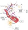
Concept drawing of the pre-metastatic niche. The primary tumor releases signals via exosomes and soluble factors to distant organs, preparing them for metastasis. Most of the distant modulation takes place in and around the microvasculature, with the endothelium being the first barrier for micrometastatic seeding. Among the most important aspects that define the pre-metastatic niche are the remodeling of the ECM, pioneering angiogenesis, the direct modulation of the endothelium and influences on the local immunological milieu.
Figure 2
Figure 2
The blood–brain barrier in the brain PMN. The blood–brain barrier (BBB) is a unique feature of the brain and is virtually the only entrance site for circulating tumor cells. As such, it has unique gatekeeping properties that are routinely modulated and hijacked by primary tumors. What little is known about the brain pre-metastatic niche overwhelmingly concerns the BBB. Dissolution of the tight junctions by immunological factors and growth factors has been shown in a pre-metastatic setting. Astrogliosis plays an important role in pro-metastatic vascular leakiness, and multiple routes lead to it; inflammation of surrounding astrocytes leads to scarring and increased BBB permeability, as does the differentiation of neural progenitor cells into astrocytes.
Figure 3
Figure 3
Advanced and experimental techniques for characterizing the pre-metastatic niche in the brain. In order to adequately describe the brain pre-metastatic niche, new and emerging techniques should be adapted to gain more insights into this elusive concept. In a spontaneous metastasis model, micro- and later oligmetastases will form in the brain. The timepoint right before the formation of the first micrometastases will represent the pre-metastatic niche most closely. Techniques such as elaborate CSF analysis for tumor cells or in vivo imaging of the brain could distinguish this timeframe. In a second phase, advanced microscopy methods combined with single-cell sequencing and spatial transcriptomics can be used to define it in more detail.

References

    1. Mattiuzzi C., Lippi G. Current Cancer Epidemiology. J. Epidemiol. Glob. Health. 2019;9:217–222. doi: 10.2991/jegh.k.191008.001. - DOI - PMC - PubMed
    1. Sacks P., Rahman M. Epidemiology of Brain Metastases. Neurosurg. Clin. 2020;31:481–488. doi: 10.1016/j.nec.2020.06.001. - DOI - PubMed
    1. Boire A., Brastianos P.K., Garzia L., Valiente M. Brain Metastasis. Nat. Rev. Cancer. 2020;20:4–11. doi: 10.1038/s41568-019-0220-y. - DOI - PubMed
    1. Kotecha R., Gondi V., Ahluwalia M.S., Brastianos P.K., Mehta M.P. Recent Advances in Managing Brain Metastasis. F1000Research. 2018;7:F1000. doi: 10.12688/f1000research.15903.1. - DOI - PMC - PubMed
    1. Valiente M., Ahluwalia M.S., Boire A., Brastianos P.K., Goldberg S.B., Lee E.Q., Le Rhun E., Preusser M., Winkler F., Soffietti R. The Evolving Landscape of Brain Metastasis. Trends Cancer. 2018;4:176–196. doi: 10.1016/j.trecan.2018.01.003. - DOI - PMC - PubMed