Review
. 2023 Jun 13;24(12):10066.
doi: 10.3390/ijms241210066.

Decipher the Immunopathological Mechanisms and Set Up Potential Therapeutic Strategies for Patients with Lupus Nephritis

Affiliations
Review

Decipher the Immunopathological Mechanisms and Set Up Potential Therapeutic Strategies for Patients with Lupus Nephritis

Chang-Youh Tsai et al. Int J Mol Sci. .

Abstract

Lupus nephritis (LN) is one of the most severe complications in patients with systemic lupus erythematosus (SLE). Traditionally, LN is regarded as an immune complex (IC) deposition disease led by dsDNA-anti-dsDNA-complement interactions in the subendothelial and/or subepithelial basement membrane of glomeruli to cause inflammation. The activated complements in the IC act as chemoattractants to chemically attract both innate and adaptive immune cells to the kidney tissues, causing inflammatory reactions. However, recent investigations have unveiled that not only the infiltrating immune-related cells, but resident kidney cells, including glomerular mesangial cells, podocytes, macrophage-like cells, tubular epithelial cells and endothelial cells, may also actively participate in the inflammatory and immunological reactions in the kidney. Furthermore, the adaptive immune cells that are infiltrated are genetically restricted to autoimmune predilection. The autoantibodies commonly found in SLE, including anti-dsDNA, are cross-reacting with not only a broad spectrum of chromatin substances, but also extracellular matrix components, including α-actinin, annexin II, laminin, collagen III and IV, and heparan sulfate proteoglycan. Besides, the glycosylation on the Fab portion of IgG anti-dsDNA antibodies can also affect the pathogenic properties of the autoantibodies in that α-2,6-sialylation alleviates, whereas fucosylation aggravates their nephritogenic activity. Some of the coexisting autoantibodies, including anti-cardiolipin, anti-C1q, anti-ribosomal P autoantibodies, may also enhance the pathogenic role of anti-dsDNA antibodies. In clinical practice, the identification of useful biomarkers for diagnosing, monitoring, and following up on LN is quite important for its treatments. The development of a more specific therapeutic strategy to target the pathogenic factors of LN is also critical. We will discuss these issues in detail in the present article.

Keywords: NLRP3 inflammasome; anti-dsDNA antibodies; cross-reactivity; lupus nephritis; renal resident cells; type I interferon.


Conflict of interest statement

The authors declare no conflict of interest.

Figures
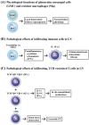
Figure 1
Figure 1
The physiological functions of the resident glomerular mesangial cells (GMCs) and macrophages (Mϕ), and the pathological activities of the infiltrating immune cells in LN. (A) The GMCs and resident macrophages can maintain normal kidney physiological functions by keeping local homeostasis and renal organogenesis. (B) The specific infiltrating macrophages with the presence of fractalkine and CD16 may produce many different cytokines/chemokines to let dendritic cells (DC) differentiate into IL-18 (+) plasmacytoid DCs (pDC), but not myeloid DCs, in the LN kidneys. These changes may lead to glomerulosclerosis and interstitial fibrosis. On the other hand, (C) the pathogenic infiltrating T cells with TCR restricted to CCR4+ CXCR3+ CD4+ and bearing TCR Vβ8+ Vβ20+ (accounting for 50%) or TCR Vβ9+Vβ14+ (accounting for 42%) produce IL-4 and IL-10 for autoantibody production. In addition, an increased CD8+T cell infiltration in the LN kidney facilitates the development of crescentic glomerulonephritis. ↑: Increase in production; ↓: Decrease in production.
Figure 2
Figure 2
The physiological functions of podocytes and the pathological basis of lupus podocytopathy. (A) The normal glomerular podocytes express many peptide molecules on the cell surface of the foot process for selectivity of glomerular filtrates in maintaining the body fluid homeostasis. (B) Defective nephrin expression on the podocyte foot process effaces the number of foot processes and suppresses the autophagic activity of podocytes. These changes may diminish the immune complex deposition in a foot process and shift it to GMCs. The shift leads to pathology, simulating a minimal-change disease or primary focal segmental glomerulonephritis in lupus patients showing the typical findings of lupus podocytopathy. ↓: Decrease in production.
Figure 3
Figure 3
The physiological and pathological functions of glomerular mesangial cells (GMCs) in patients with LN. After activation or damage by the immune complex, a GMC secretes proinflammatory cytokines, IL-1β, IL-6, and IFN-I, to accelerate the inflammation change within the kidney. On the other hand, an extracellular matrix (ECM) is also accumulated to enhance tissue fibrosis. Both of the changes ultimately result in LN. ↑: Increase in production.
Figure 4
Figure 4
The intricate interactions among a systemic autoimmune reaction, infiltrating immune-related cells and the renal resident cells in inducing lupus nephritis. Not only cytokines, chemokines, and growth factors, but also the formation of a tertiary lymphoid structure (TLS) containing plasma cells with somatic hypermutation is involved in the in situ generation of autoreactive antibodies. In addition, the T-cell-rich zone in a TLS contains Th1 phenotypes, which are reactive with kidney-specific antigens, mature dendritic cells (DCs), Th17 cells, and CD4+ Treg to destroy the kidney resident cells. These factors in the tissue altogether induce inflammation, cell proliferation, cell damage, and fibrosis—commonly found in the progression of lupus nephritis. ↑: Increase in production.
Figure 5
Figure 5
Comparison of different chemokines and cytokines in the induction of murine (left panel) and human (right panel) lupus nephritis. In human LN, both B cell and inflammatory cell activation accelerate the progression. (Regarding the abbreviations, please refer to the abbreviation list at the end of the text).
Figure 6
Figure 6
The differences of NLRP3 inflammasome activation between normal human innate immune cells and the renal podocytes in patients with SLE. (A) The binding of PAMP or DAMP molecules to NLR activates RIP3 and then NLRP3 inflammasome. The inflammasome activation increases IL-1β and IL-18 production to enhance inflammatory responses and cell necroptosis. (B) The podocytes in SLE kidney not only repress NLR, but autonomously express excessive CD36, a class B scavenger receptor, to bind with PAMP and DAMP. This excessive CD36 expression in SLE may facilitate spontaneous RIP3 activation and subsequent NLRP3 inflammasome activation, as well as impeding autophagy activity, and eventually results in necroptosis of podocytes and lupus nephritis. ↑: Increase in production; ↓: Decrease in production.
Figure 7
Figure 7
The molecular basis for the effects of pathogenic dsDNA antibodies on somatic and different renal resident cells—including podocytes, mesangial cells, and epithelial cells—by inducing cell damage, inflammation, and tissue fibrosis. (A) Anti-dsDNA antibodies cross-react with a human acidic ribosomal P protein expressed on different somatic cells (liver, spleen, or MC) to elicit cell apoptosis. (B) Anti-dsDNA antibodies cross-react with α-actinin, an F-actin binding protein present in the cell–cell and cell–matrix contact sites for maintaining cell morphology and integrity. The binding of anti-dsDNA with α-actinin on podocyte cell surface causes disfiguration dysmotility and even cell death. (C) The binding of anti-dsDNA with acidic ribosomal P expressed on glomerular mesangial cell surface induce an IL-6 expression and inflammation. (D) The anti-dsDNA antibodies may also bind to nonspecific IgG-Fcγ receptors on renal epithelial cells to stimulate excretion of chemokines and other molecules, eventually resulting in chronic inflammation/tissue fibrosis, in addition to the aforementioned cross-reactivity with the surface-expressed molecules. Epi: renal epithelial cell. ↑: Increase in production.

References

    1. Hoover P.J., Costenbader K.H. Insights into the epidemiology and management of lupus nephritis from the US rheumatologist’s perspective. Kidney Int. 2016;90:487–492. doi: 10.1016/j.kint.2016.03.042. - DOI - PMC - PubMed
    1. Hanly J.G., O’Keeffe A.G., Su L., Urowitz M.B., Romero-Diaz J., Gordon C., Bae S.-C., Bernatsky S., Clarke A.E., Wallace D.J., et al. The frequency and outcome of lupus nephritis: Results from an international inception cohort study. Rheumatology. 2016;55:252–262. doi: 10.1093/rheumatology/kev311. - DOI - PMC - PubMed
    1. Weening J.J., D’Agati V.D., Schwartz M.M., Seshan S.V., Alpers C.E., Appel G.B., Balow J.E., Bruijn J.A., Cook T., Ferrario F., et al. The classification of glomerulonephritis in systemic lupus erythematosus revisited. Kidney Int. 2004;65:521–530. doi: 10.1111/j.1523-1755.2004.00443.x. - DOI - PubMed
    1. Moroni G., Depetri F., Ponticelli C. Lupus nephritis: When and how often to biopsy and what does it mean? J. Autoimmun. 2016;74:27–40. doi: 10.1016/j.jaut.2016.06.006. - DOI - PubMed
    1. Bastian H.M., Roseman J.M., McGwin G., Jr., Alarcón G.S., Friedman A.W., Fessler B.J., Baethge B.A., Reveille J.D., LUMINA Study Group Systemic lupus erythematosus in three ethnic groups. XII. Risk factors for lupus nephritis after diagnosis. Lupus. 2002;11:152–160. doi: 10.1191/0961203302lu158oa. - DOI - PubMed