. 2023 Jun 15;24(12):10177.
doi: 10.3390/ijms241210177.

New Perspectives in Dried Blood Spot Biomarkers for Lysosomal Storage Diseases

Affiliations

New Perspectives in Dried Blood Spot Biomarkers for Lysosomal Storage Diseases

Justyna Spiewak et al. Int J Mol Sci. .

Abstract

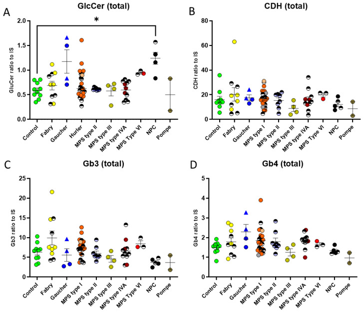
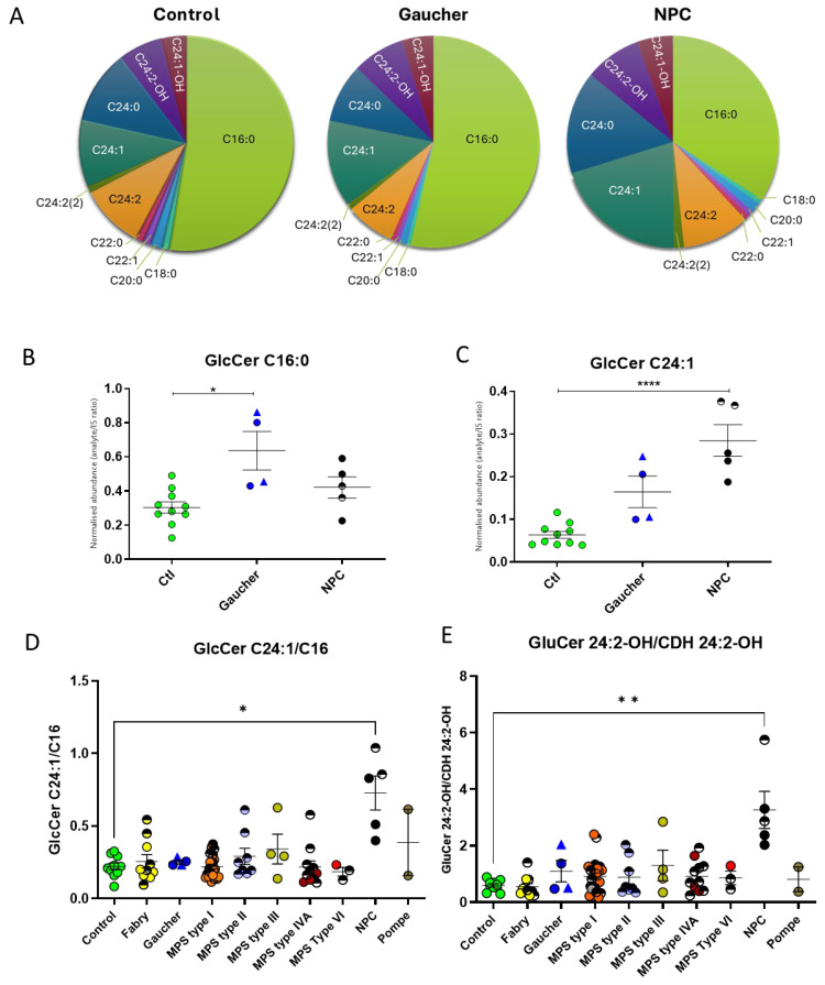
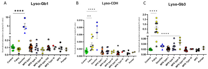
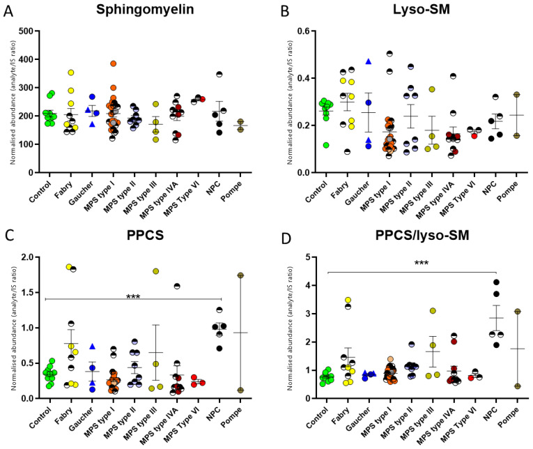
Dried blood spots (DBSs) biomarkers are convenient for monitoring for specific lysosomal storage diseases (LSDs), but they could have relevance for other LSDs. To determine the specificity and utility of glycosphingolipidoses biomarkers against other LSDs, we applied a multiplexed lipid liquid chromatography tandem mass spectrometry assay to a DBS cohort of healthy controls (n = 10) and Gaucher (n = 4), Fabry (n = 10), Pompe (n = 2), mucopolysaccharidosis types I-VI (n = 52), and Niemann-Pick disease type C (NPC) (n = 5) patients. We observed no complete disease specificity for any of the markers tested. However, comparison among the different LSDs highlighted new applications and perspectives of the existing biomarkers. We observed elevations in glucosylceramide isoforms in the NPC and Gaucher patients relative to the controls. In NPC, there was a greater proportion of C24 isoforms, giving a specificity of 96-97% for NPC, higher than 92% for the NPC biomarker N-palmitoyl-O-phosphocholineserine ratio to lyso-sphingomyelin. We also observed significantly elevated levels of lyso-dihexosylceramide in Gaucher and Fabry disease as well as elevated lyso-globotriaosylceramide (Lyso-Gb3) in Gaucher disease and the neuronopathic forms of Mucopolysaccharidoses. In conclusion, DBS glucosylceramide isoform profiling has increased the specificity for the detection of NPC, thereby improving diagnostic accuracy. Low levels of lyso-lipids can be observed in other LSDs, which may have implications in their disease pathogenesis.

Keywords: Fabry disease; Gaucher disease; Niemann–Pick C disease; biomarker; dried blood spot; glycosphingolipid; mucopolysaccharidoses.


Conflict of interest statement

The authors declare no conflict of interest.

Figures

Figure 1
Figure 1
The glycosphingolipid degradation pathway, illustrating known enzyme defects and associated disorders (red text). Lipids that are included in the assay are indicated by *. PPCS presented individually, as the associated pathway is unknown.
Figure 2
Figure 2
Dried blood spot glycosphingolipid levels in a cohort of patients with lysosomal storage diseases. The four main GSL species detectable in DBS are shown. (A) GlcCer—only significantly different to the controls for NPC, and therefore useful as a biomarker. (B) CDH—no significant changes observed. (C) Gb3 CDH—no significant changes observed. (D) GB4 CDH—no significant changes observed. Patients in treatment are represented by half shaded circles. Gaucher type I is represented by a triangle symbol, and type III, by a circle. The untreated MPS type I group is depicted as follows: Hurler in orange, Hurler–Scheie in grey, and Scheie in pink. Significance determined by Kruskal–Wallis non-parametric test. * means significance of p ≤ 0.05.
Figure 3
Figure 3
Glucosylceramide isoforms in dried blood spots from patients with lysosomal storage diseases. (A) A summary pie chart of the mean values of each isoform in the control, Gaucher, and NPC disease, showing a differing isoform profile in NPC disease. (B) The C16 isoform in Gaucher and NPC DBS in comparison with control levels. (C) The C24:1 isoform in Gaucher and NPC DBS in comparison with control levels. (D) Ratio of GlcCer C24:1/C16 isoforms in a cohort of patients with LSDs, showing that NPC has the greatest ratio difference relative to controls. (E) Ratio of GlcCer 24:2-OH/CDH 24:2-OH isoforms, showing the best specificity for NPC. Patients on treatment are represented by half-shaded circles. Gaucher type I is indicated by a triangle symbol, and type III, by a circle. The untreated MPS type I group circles are coloured as follows: Hurler as orange, Hurler–Scheie as grey, and Scheie as pink. **** means significance of p ≤ 0.0001; ** means significance of p ≤ 0.01; * means significance of p ≤ 0.05.
Figure 4
Figure 4
Lyso-glycosphingolipid dried-blood-spot analysis of samples from a cohort of patients with lysosomal storage diseases. (A) The Gaucher disease group has the highest levels of lyso-Gb1. Low-level changes in lyso-Gb1 in other LSDs were observed. (B) Small increases in the levels of lyso-CDH were seen in the Fabry and Gaucher samples. (C) The Fabry disease group has the highest lyso-Gb3 levels. Much smaller changes were observed for the other LSDs patients on treatment, represented by half-shaded circles. Gaucher type I samples are represented by a triangle symbol, and type III, by a circle. The untreated MPS type I group circles are coloured as follows: Hurler is orange, Hurler–Scheie is grey, and Scheie is pink. **** means significance of p ≤ 0.0001; ** means significance of p ≤ 0.01.
Figure 5
Figure 5
Levels of NPC biomarkers in a cohort of patients with lysosomal storage diseases. (A) Levels of total sphingomyelin in the LSD groups are similar to those seen in control samples. (B) Lyso-sphingomyelin levels are similar to those seen in the control cohort, with some outlier patients. (C) Total PPCS is significantly elevated in NPC but shows outliers in other disorders. (D) Ratioing of PPCS to lyso-sphingomyelin improves specificity for NPC relative to other LSDs. Patients on treatment are represented by half-shaded circles. Gaucher type I is indicated by a triangle symbol, and type III, by a circle. The untreated MPS type I group circles are coloured as follows: Hurler in orange, Hurler–Scheie in grey, and Scheie in pink. *** significance of p ≤ 0.001.

References

    1. Papandreou A., Doykov I., Spiewak J., Komarov N., Habermann S., Kurian M.A., Mills P.B., Mills K., Gissen P., Heywood W.E., et al. Niemann-Pick type C disease as proof-of-concept for intelligent biomarker panel selection in neurometabolic disorders. Dev. Med. Child Neurol. 2022;64:1539–1546. doi: 10.1111/dmcn.15334. - DOI - PMC - PubMed
    1. Dinur T., Bauer P., Beetz C., Kramp G., Cozma C., Iurascu M.I., Becker-Cohen M., Istaiti M., Rolfs A., Zimran A., et al. Gaucher Disease Diagnosis Using Lyso-Gb1 on Dry Blood Spot Samples: Time to Change the Paradigm? Int. J. Mol. Sci. 2022;23:1627. doi: 10.3390/ijms23031627. - DOI - PMC - PubMed
    1. Malvagia S., Ferri L., Della Bona M., Borsini W., Cirami C.L., Dervishi E., Feriozzi S., Gasperini S., Motta S., Mignani R., et al. Multicenter evaluation of use of dried blood spot compared to conventional plasma in measurements of globotriaosylsphingosine (LysoGb3) concentration in 104 Fabry patients. Clin. Chem. Lab. Med. 2021;59:1516–1526. doi: 10.1515/cclm-2021-0316. - DOI - PubMed
    1. Khaledi H., Gelb M.H. Tandem Mass Spectrometry Enzyme Assays for Multiplex Detection of 10-Mucopolysaccharidoses in Dried Blood Spots and Fibroblasts. Anal. Chem. 2020;92:11721–11727. doi: 10.1021/acs.analchem.0c01750. - DOI - PMC - PubMed
    1. Young E., Mills K., Morris P., Vellodi A., Lee P., Waldek S., Winchester B. Is globotriaosylceramide a useful biomarker in Fabry disease? Acta Paed. 2005;94:51–54; discussion 37–38. doi: 10.1080/08035320510028111. - DOI - PubMed

LinkOut - more resources