. 2023 Jun 16;24(12):10204.
doi: 10.3390/ijms241210204.

Bromotyrosine-Derived Metabolites from a Marine Sponge Inhibit Pseudomonas aeruginosa Biofilms

Affiliations

Bromotyrosine-Derived Metabolites from a Marine Sponge Inhibit Pseudomonas aeruginosa Biofilms

Tam M T Tran et al. Int J Mol Sci. .

Abstract

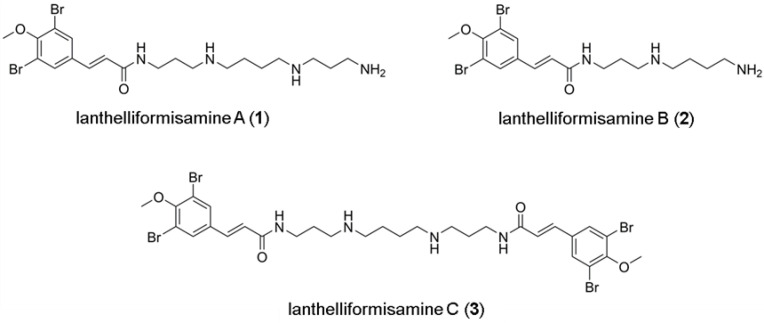
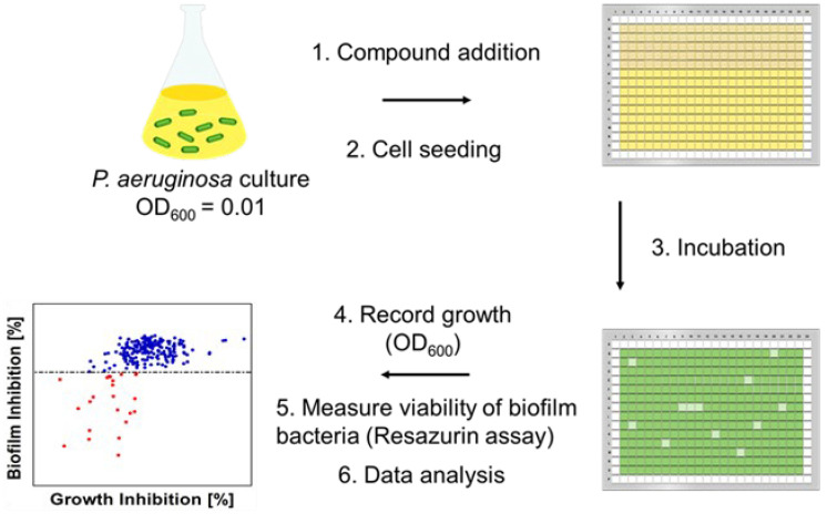
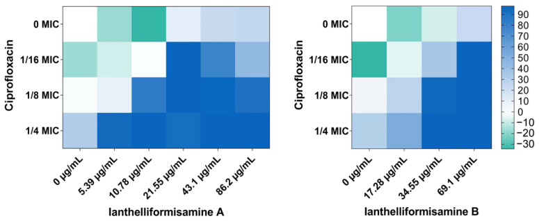
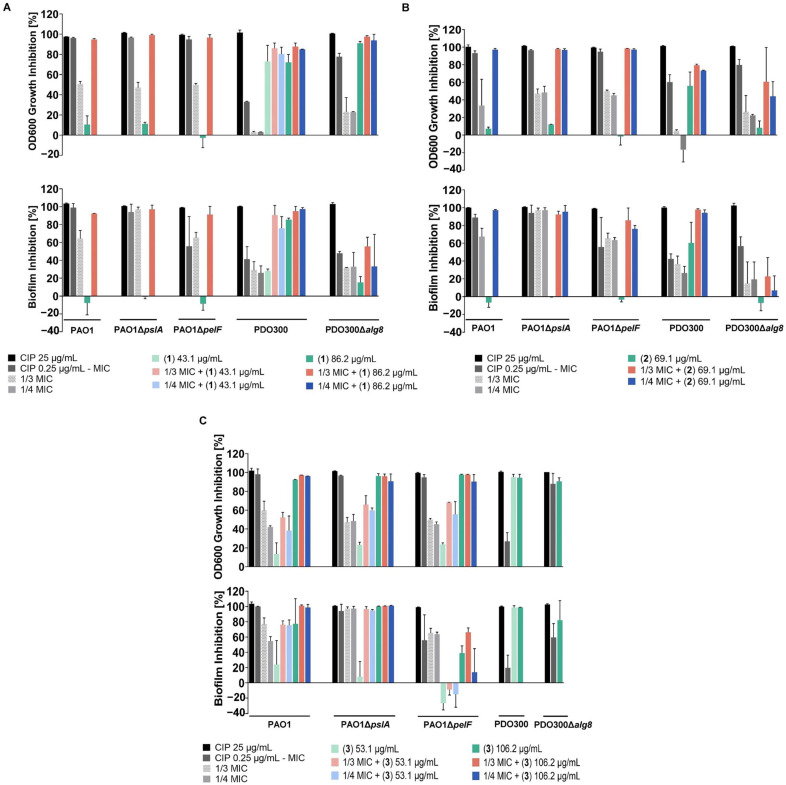
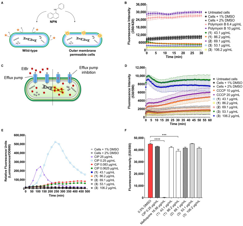
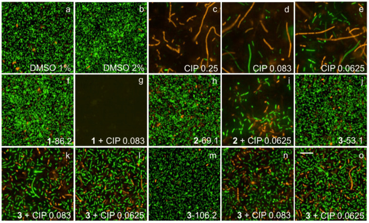
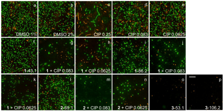
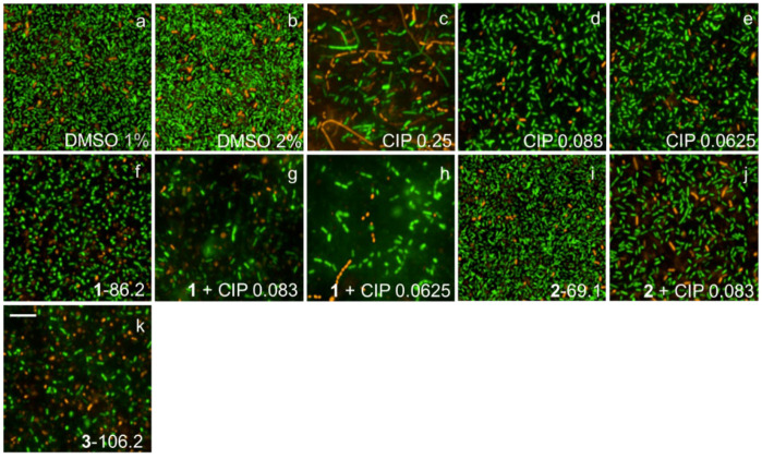
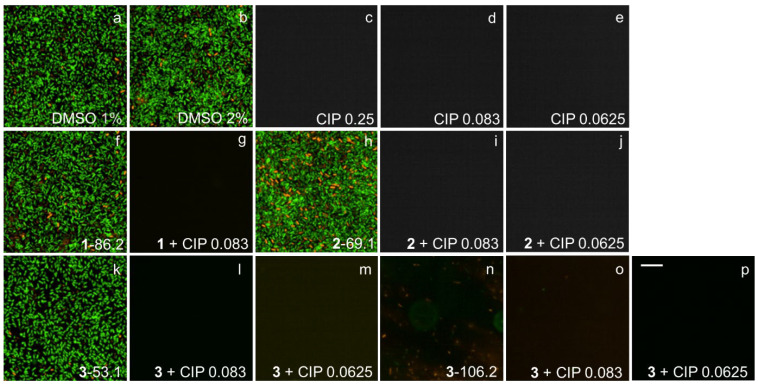
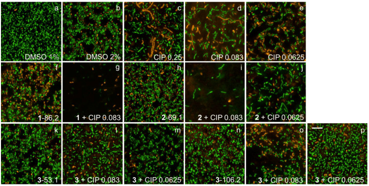
Pseudomonas aeruginosa forms stable biofilms, providing a major barrier for multiple classes of antibiotics and severely impairing treatment of infected patients. The biofilm matrix of this Gram-negative bacterium is primarily composed of three major exopolysaccharides: alginate, Psl, and Pel. Here, we studied the antibiofilm properties of sponge-derived natural products ianthelliformisamines A-C and their combinations with clinically used antibiotics. Wild-type P. aeruginosa strain and its isogenic exopolysaccharide-deficient mutants were employed to determine the interference of the compounds with biofilm matrix components. We identified that ianthelliformisamines A and B worked synergistically with ciprofloxacin to kill planktonic and biofilm cells. Ianthelliformisamines A and B reduced the minimum inhibitory concentration (MIC) of ciprofloxacin to 1/3 and 1/4 MICs, respectively. In contrast, ianthelliformisamine C (MIC = 53.1 µg/mL) alone exhibited bactericidal effects dose-dependently on both free-living and biofilm populations of wild-type PAO1, PAO1ΔpslA (Psl deficient), PDO300 (alginate overproducing and mimicking clinical isolates), and PDO300Δalg8 (alginate deficient). Interestingly, the biofilm of the clinically relevant mucoid variant PDO300 was more susceptible to ianthelliformisamine C than strains with impaired polysaccharide synthesis. Ianthelliformisamines exhibited low cytotoxicity towards HEK293 cells in the resazurin viability assay. Mechanism of action studies showed that ianthelliformisamine C inhibited the efflux pump of P. aeruginosa. Metabolic stability analyses indicated that ianthelliformisamine C is stable and ianthelliformisamines A and B are rapidly degraded. Overall, these findings suggest that the ianthelliformisamine chemotype could be a promising candidate for the treatment of P. aeruginosa biofilms.

Keywords: Pseudomonas aeruginosa; alkaloid; biofilms; bromotyrosine; ciprofloxacin; cytotoxicity; ianthelliformisamines; in vitro metabolism; natural product; sponge.


Conflict of interest statement

The authors declare no competing interest.

Figures

Figure 1
Figure 1
Chemical structures of ianthelliformisamines A–C.
Figure 2
Figure 2
Biofilm assays to identify compounds that prevent biofilm formation. Biofilm inhibition assay. P. aeruginosa cultures were seeded into 384-well plates after dispensing compounds and antibiotics. The plates were incubated for 24 h at 37 °C followed by recording bacterial growth (OD600) and measuring the viability of biofilm bacteria using resazurin.
Figure 3
Figure 3
Ianthelliformisamine A and ianthelliformisamine B synergized with ciprofloxacin (CIP). Heat plots showing growth inhibition of planktonic PAO1 in the presence of compound and ciprofloxacin. Percent of growth inhibition is illustrated with different colors, where green represents growth stimulation and dark blue 100% inhibition.
Figure 4
Figure 4
Ianthelliformisamines A–C (13) combined with ciprofloxacin resulted in decreased cell growth and biofilm formation. To assess interference with biofilm matrix components, wild-type and different isogenic mutant strains of P. aeruginosa PAO1, PAO1ΔpslA, PAO1ΔpelF, PDO300, and PDO300Δalg8 were treated with 1 (A), 2 (B), 3 (C), or ciprofloxacin, alone or in combination for 24 h. Cell growth (OD600) was monitored, and the resazurin assay was performed to determine the viability of biofilm bacteria. A culture with matched percentages of DMSO was used as the negative (untreated) controls. The experiment was conducted in triplicate. The results are percentage means and standard deviation (SD) of two independent experiments. Abbreviation: CIP, ciprofloxacin.
Figure 5
Figure 5
(A) The NPN dye is a lipophilic molecule that is used to determine permeability changes in bacteria. Such molecules are weakly fluorescent in an aqueous environment and impermeable to the cell with an intact outer membrane. However, once the membrane is damaged, NPN penetrates the cells and binds to the phospholipid layer, giving rise to pronounced fluorescence. (B) Wild-type PAO1 cells were treated with 100 µM CCCP prior to exposure to test compounds at different concentrations. The permeability changes in the outer membrane were assessed by monitoring the fluorescence of NPN for 30 min. Cells treated with DMSO solvent were included as the negative control. Polymyxin B was used as a positive control. (C) Ethidium bromide (EtBr) is a common substrate of the resistance-nodulation-cell division (RND) pump, which is used to measure the amount of intracellular accumulation, as it only fluoresces when bound to DNA. (D) The accumulation of EtBr in the presence of CCCP and test compounds was monitored for 60 min. Cells treated with DMSO solvent were included as the negative control. CCCP was used as a positive control. (E) Test compounds do not inhibit DNA synthesis. The reporter strain MDM-623 harboring the promoter region from PA0614 fused to P. luminescens luxCDABE operon, a promoter-luciferase reporter gene, constructed to respond to DNA damage in general. The optical density OD600 and the kinetics of luminescence of the reporter strain were monitored for 8 h. Cells treated with DMSO solvent were included as the negative control, while ciprofloxacin at different concentrations was used as a positive control. (F) HEK293 resazurin viability assay. HEK293 cells were incubated with test compounds to evaluate cytotoxicity by resazurin assay. After 24 h of incubation, resazurin was dispensed into the wells, and the plate was further incubated for 5 h at 37 °C. Cells treated with DMSO solvent were included as the negative control. Mefloquine was used as a positive control. All experiments were performed in triplicate and repeated twice. The data are the means and SDs of two independent experiments. A non-parametric one-way ANOVA followed by Dunnett’s multiple comparison post-hoc test was carried out to determine statistical significance between each treatment and the negative control (0.5% DMSO).; ***, p < 0.001; ****, p < 0.0001. Abbreviation: CIP, ciprofloxacin.
Figure 6
Figure 6
Representative fluorescence images of treated and untreated PAO1 biofilms. Cells treated with ciprofloxacin and test compounds 13 were stained with SYTO9 (green) and propidium iodide (red). Ciprofloxacin generated long, filamentous phenotypes as well as small, round spheroplasts. Biofilms challenged with 1 and 2 remained unaffected, while that with 3 was slightly reduced. Complete biofilm inhibition was observed with 1 + 1/3 MIC. Abbreviations: 13 (ianthelliformisamines A–C), CIP (ciprofloxacin). Cells treated with (a) DMSO 1%, (b) DMSO 2%, (c) ciprofloxacin at 1 × MIC, (d) ciprofloxacin at 1/3 MIC, (e) ciprofloxacin at 1/4 MIC, (f) 1 at 86.2 µg/mL, (g) 1 86.2 µg/mL + CIP 1/3 MIC, (h) 2 at 69.1 µg/mL, (i) 2 69.1 µg/mL + CIP 1/4 MIC, (j) 3 at 53.15 µg/mL, (k) 3 53.1 µg/mL + CIP 1/3 MIC, (l) 3 53.1 µg/mL + CIP 1/4 MIC, (m) 3 at 106.2 µg/mL, (n) 3 106.2 µg/mL + CIP 1/3 MIC, and (o) 3 106.2 µg/mL + CIP 1/4 MIC. Scale bar 10 µM.
Figure 7
Figure 7
Representative fluorescence images of treated and untreated PDO300 biofilms. Cells treated with ciprofloxacin and test compounds 13 were stained with SYTO9 (green) and propidium iodide (red). Ciprofloxacin generated long, filamentous phenotypes as well as small, round spheroplasts. Biofilms challenged with 1 and 2 slightly reduced, while those challenged with 3 were completely eradicated. Abbreviations: 13 (ianthelliformisamines A–C), CIP (ciprofloxacin). Cells treated with (a) DMSO 1%, (b) DMSO 2%, (c) ciprofloxacin at 1 × MIC, (d) ciprofloxacin at 1/3 MIC, (e) ciprofloxacin at 1/4 MIC, (f) 1 at 43.1 µg/mL, (g) 1 43.1 µg/mL + CIP 1/3 MIC, (h) 1 43.1 µg/mL + CIP 1/4 MIC, (i) 1 at 86.2 µg/mL, (j) 1 86.2 µg/mL + CIP 1/3 MIC, (k) 1 86.2 µg/mL + CIP 1/4 MIC, (l) 2 at 69.1 µg/mL, (m) 2 69.1 µg/mL + CIP 1/3 MIC, (n) 2 69.1 µg/mL + CIP 1/4 MIC, (o) 3 at 53.1 µg/mL, and (p) 3 at 106.2 µg/mL. Scale bar 10 µM.
Figure 8
Figure 8
Representative fluorescence images of treated and untreated PDO300Δalg8 biofilms. Cells treated with ciprofloxacin and test compounds 13 were stained with SYTO9 (green) and propidium iodide (red). Ciprofloxacin generated long, filamentous phenotypes as well as small, round spheroplasts. Biofilms challenged with 13 were inhibited at different levels. Abbreviations: 13 (ianthelliformisamines A–C), CIP (ciprofloxacin). Cells treated with (a) DMSO 1%, (b) DMSO 2%, (c) ciprofloxacin at 1 × MIC, (d) ciprofloxacin at 1/3 MIC, (e) ciprofloxacin at 1/4 MIC, (f) 1 at 86.2 µg/mL, (g) 1 86.2 µg/mL + CIP 1/3 MIC, (h) 1 86.2 µg/mL + CIP 1/4 MIC, (i) 2 at 69.1 µg/mL, (j) 2 69.1 µg/mL + CIP 1/3 MIC, and (k) 3 at 106.2 µg/mL. Scale bar 10 µM.
Figure 9
Figure 9
Representative fluorescence images of treated and untreated PAO1ΔpslA biofilms. Cells treated with ciprofloxacin and test compounds 13 were stained with SYTO9 (green) and propidium iodide (red). PAO1ΔpslA was unable to form biofilms in the presence of ciprofloxacin. Biofilms challenged with 13 remained unaffected. Abbreviations: 13 (ianthelliformisamines A–C), CIP (ciprofloxacin). Cells treated with (a) DMSO 1%, (b) DMSO 2%, (c) ciprofloxacin at 1 × MIC, (d) ciprofloxacin at 1/3 MIC, (e) ciprofloxacin at 1/4 MIC, (f) 1 at 86.2 µg/mL, (g) +1 86.2 µg/mL + CIP 1/3 MIC, (h) 2 at 69.1 µg/mL, (i) 2 69.1 µg/mL + CIP 1/3 MIC, (j) 2 69.1 µg/mL + CIP 1/4 MIC, (k) 3 at 53.1 µg/mL, (l) 3 53.1 µg/mL + CIP 1/3 MIC, (m) 3 53.1 µg/mL + CIP 1/4 MIC, (n) 3 at 106.2 µg/mL, (o) 3 106.2 + CIP 1/3 MIC µg/mL, and (p) 3 106.2 + CIP 1/4 MIC µg/mL. Scale bar 10 µM.
Figure 10
Figure 10
Representative fluorescence images of treated and untreated PAO1ΔpelF biofilms. Cells treated with ciprofloxacin and test compounds 13 were stained with SYTO9 (green) and propidium iodide (red). Ciprofloxacin generated long, filamentous phenotypes as well as small, round spheroplasts. Biofilms challenged with 13 remained unaffected. Abbreviations: 13 (ianthelliformisamines A–C), CIP (ciprofloxacin). Cells treated with (a) DMSO 1%, (b) DMSO 2%, (c) ciprofloxacin at 1 × MIC, (d) ciprofloxacin at 1/3 MIC, (e) ciprofloxacin at 1/4 MIC, (f) 1 at 86.2 µg/mL, (g) 1 86.2 µg/mL + CIP 1/3 MIC, (h) 2 at 69.1 µg/mL, (i) 2 69.1 µg/mL + CIP 1/3 MIC, (j) 2 69.1 µg/mL + CIP 1/4 MIC, (k) 3 at 53.1 µg/mL, (l) 3 53.1 µg/mL + CIP 1/3 MIC, (m) 3 53.1 µg/mL + CIP 1/4 MIC, (n) 3 at 106.2 µg/mL, (o) 3 106.2 µg/mL + CIP 1/3 MIC, and (p) 3 106.2 µg/mL + CIP 1/4 MIC. Scale bar 10 µM.

References

    1. Moradali M.F., Ghods S., Rehm B.H.A. Pseudomonas aeruginosa Lifestyle: A Paradigm for Adaptation, Survival, and Persistence. Front. Cell. Infect. Microbiol. 2017;7:39. doi: 10.3389/fcimb.2017.00039. - DOI - PMC - PubMed
    1. Moradali M.F., Rehm B.H.A. Bacterial biopolymers: From pathogenesis to advanced materials. Nat. Rev. Microbiol. 2020;18:195–210. doi: 10.1038/s41579-019-0313-3. - DOI - PMC - PubMed
    1. Penesyan A., Nagy S.S., Kjelleberg S., Gillings M.R., Paulsen I.T. Rapid microevolution of biofilm cells in response to antibiotics. NPJ Biofilms Microbiomes. 2019;5:34. doi: 10.1038/s41522-019-0108-3. - DOI - PMC - PubMed
    1. Ciofu O., Tolker-Nielsen T. Tolerance and Resistance of Pseudomonas aeruginosa Biofilms to Antimicrobial Agents-How P. aeruginosa Can Escape Antibiotics. Front. Microbiol. 2019;10:9132019. doi: 10.3389/fmicb.2019.00913. - DOI - PMC - PubMed
    1. Ceri H., Olson M.E., Stremick C., Read R.R., Morck D., Bure A. The Calgary Biofilm Device: New technology for rapid determination of antibiotic susceptibilities of bacterial biofilms. J. Clin. Microbiol. 1999;37:1771–1776. doi: 10.1128/JCM.37.6.1771-1776.1999. - DOI - PMC - PubMed

LinkOut - more resources