. 2023 Jun 20;24(12):10392.
doi: 10.3390/ijms241210392.

Impact of Manganese and Chromate on Specific DNA Double-Strand Break Repair Pathways

Affiliations

Impact of Manganese and Chromate on Specific DNA Double-Strand Break Repair Pathways

Vivien M M Haberland et al. Int J Mol Sci. .

Abstract

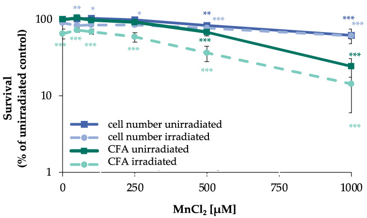
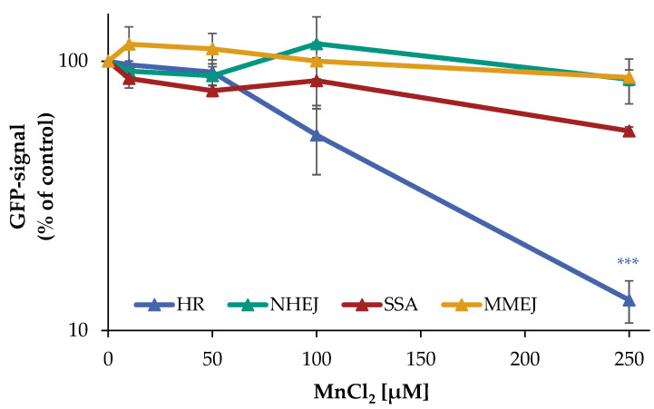
Manganese is an essential trace element; nevertheless, on conditions of overload, it becomes toxic, with neurotoxicity being the main concern. Chromate is a well-known human carcinogen. The underlying mechanisms seem to be oxidative stress as well as direct DNA damage in the case of chromate, but also interactions with DNA repair systems in both cases. However, the impact of manganese and chromate on DNA double-strand break (DSB) repair pathways is largely unknown. In the present study, we examined the induction of DSB as well as the effect on specific DNA DSB repair mechanisms, namely homologous recombination (HR), non-homologous end joining (NHEJ), single strand annealing (SSA), and microhomology-mediated end joining (MMEJ). We applied DSB repair pathway-specific reporter cell lines, pulsed field gel electrophoresis as well as gene expression analysis, and investigated the binding of specific DNA repair proteins via immunoflourescence. While manganese did not seem to induce DNA DSB and had no impact on NHEJ and MMEJ, HR and SSA were inhibited. In the case of chromate, the induction of DSB was further supported. Regarding DSB repair, no inhibition was seen in the case of NHEJ and SSA, but HR was diminished and MMEJ was activated in a pronounced manner. The results indicate a specific inhibition of error-free HR by manganese and chromate, with a shift towards error-prone DSB repair mechanisms in both cases. These observations suggest the induction of genomic instability and may explain the microsatellite instability involved in chromate-induced carcinogenicity.

Keywords: BRCA1; DNA DSB repair; HR; MMEJ; NHEJ; RAD51; RAD54; SSA; U2OS reporter-assay; chromate; manganese.


Conflict of interest statement

The authors declare no conflict of interest.

Figures

Figure 1
Figure 1
Impact of manganese on cell number and CFA in HeLa S3 cells after irradiation. Cells were pre-incubated with MnCl2 for 24 h, irradiated with 1 Gy and post-incubated in the presence of MnCl2 for 8 h. Cells were subsequently trypsinized, counted (cell number), and reseeded for assessment of colony forming ability (CFA). Results are normalized to cell number or CFA of the untreated control. Shown are mean values of three independent experiments performed each with double determination ± SD. Statistical analysis between manganese treatment and corresponding controls (* p ≤ 0.05, ** p ≤ 0.01, *** p ≤ 0.001) were performed by one-way ANOVA with post hoc Dunnett T.
Figure 2
Figure 2
Impact of manganese on the repair of IR-induced DSB measured by PFGE. HeLa S3 cells were pre-incubated with MnCl2 for 24 h and irradiated with 80 Gy. Cells were harvested directly afterwards or post-incubated in the presence of MnCl2 for further 8 h. (A) DNA fragments were separated by gel electrophoresis. Shown is one representative gel from three determinations. (B) The fraction of DNA released (FDR) value was quantified, describing the ratio of DNA fraction migrated in the gel to the total DNA content as an indicator for DNA DSB. Shown are mean values of three independent experiments, each performed in duplicate ± SD. Statistical analysis between the respective treatments and controls (* p ≤ 0.05, ** p ≤ 0.01) were performed by one-way ANOVA with post hoc Dunnett T.
Figure 3
Figure 3
Impact of manganese on the repair of DSB measured by U2OS reporter-assay. U2OS cells were transfected with ISce-I for 6 h. After removing the transfection cocktail, the cells were incubated with MnCl2 for 66 h. Cells were subsequently trypsinized, harvested, and GFP-active cells were quantified via flow cytometry. The results are normalized to the transfected control. Shown are mean values of three independent experiments performed in double determination ± SD. For each treatment, 50,000 events were counted. Statistical analysis between manganese treatment and corresponding controls (*** p ≤ 0.001) were performed by one-way ANOVA with post hoc Dunnett T.
Figure 4
Figure 4
RAD51 and RAD54 foci formation after manganese treatment and irradiation with 1 Gy in HeLa S3 cells. Cells were pre-incubated with MnCl2 for 24 h or 64 h (A,C), irradiated with 1 Gy (B,D) and post-incubated in the presence of MnCl2 for 8 h. Cells were stained against RAD51 (A,B) or RAD54 (C,D). G2 cells were identified via CENP-F staining. Shown are unirradiated (A,C) and irradiated (B,D) cells. The data represent mean values of three independent experiments performed in double determination ± SD. For each time point and treatment, respective foci were counted in 40 G2 phase cells. Statistical analysis between manganese treatment and corresponding controls (* p ≤ 0.05, ** p ≤ 0.01, *** p ≤ 0.001) were performed by one-way ANOVA with post hoc Dunnett T. Pictures displaying foci formation are shown in Supplementary Figures S1 and S2.
Figure 5
Figure 5
Overview of the impact of manganese on HeLa S3 cells using a high-throughput RT-qPCR. Cells were pre-incubated with MnCl2 for 24 h, irradiated with 1 Gy and post-incubated in the presence of MnCl2 for 8 h. Genes have been clustered into groups associated with DNA damage response and oxidative stress response. Displayed are the log2-fold changes of relative gene expression as a heatmap. Red represents an enhanced expression and blue represents a down-regulation of different genes. Shown are the mean values of at least three independently conducted experiments.
Figure 6
Figure 6
Impact of Cr(VI) on cell number and CFA of HeLa S3 cells. Cells were incubated with K2Cr2O7 for 32 h or 72 h. Cells were subsequently trypsinized, counted (cell number) and reseeded for assessment of colony forming ability (CFA). Results are normalized to cell number or CFA of the untreated control. Shown are mean values of three independent experiments performed in double determination ± SD. Statistical analysis between chromate treatment and corresponding controls (* p ≤ 0.05, ** p ≤ 0.01, *** p ≤ 0.001) were performed by one-way ANOVA with post hoc Dunnett T.
Figure 7
Figure 7
Impact of Cr(VI) on different DSB repair pathways determined by U2OS reporter-assay. U2OS cells were transfected with ISce-I for 6 h. After removing the transfection cocktail the cells were incubated with K2Cr2O7 for 66 h, to obtain a maximum GFP signal. Cells were subsequently trypsinized, harvested, and GFP-active cells were quantified via flow cytometry. The results are normalized to the transfected control. Shown are mean values of three independent experiments performed with double determinations ± SD. For each treatment 50,000 events were counted. Statistical analysis between chromate treatment and corresponding controls were performed by one-way ANOVA with post hoc Dunnett T.
Figure 8
Figure 8
BRCA1, RAD51, and RAD54 foci formation after Cr(VI) treatment and irradiation with 1 Gy in HeLa S3 cells. Cells were pre-incubated with K2Cr2O7 for 24 h or 64 h (A,C,E), irradiated with 1 Gy (B,D,F), and post-incubated in the presence of K2Cr2O7 for 8 h. Cells were stained against BRCA1 (A,B), RAD51 (C,D), and RAD54 (E,F). G2 cells were identified via CENP-F staining. Shown are unirradiated (A,C,E) and irradiated (B,D,F) cells. The data represent mean values of three independent experiments performed in double determination ± SD. For each time point and treatment, foci were counted in 40 G2 phase cells. Statistical analysis between Cr(VI) treatment and corresponding controls (* p ≤ 0.05, ** p ≤ 0.01, *** p ≤ 0.001) were performed by one-way ANOVA with post hoc Dunnett T. Pictures displaying foci formation are shown in Supplementary Figures S3–S5.

References

    1. Chen P., Bornhorst J., Aschner M. Manganese metabolism in humans. Front. Biosci. (Landmark Ed.) 2018;23:1655–1679. doi: 10.2741/4665. - DOI - PubMed
    1. Peres T.V., Schettinger M.R., Chen P., Carvalho F., Avila D.S., Bowman A.B., Aschner M. “Manganese-induced neurotoxicity: A review of its behavioral consequences and neuroprotective strategies”. BMC Pharmacol. Toxicol. 2016;17:57. doi: 10.1186/s40360-016-0099-0. - DOI - PMC - PubMed
    1. Bornhorst J., Ebert F., Hartwig A., Michalke B., Schwerdtle T. Manganese inhibits poly(ADP-ribosyl)ation in human cells: A possible mechanism behind manganese-induced toxicity? J. Environ. Monit. 2010;12:2062–2069. doi: 10.1039/c0em00252f. - DOI - PubMed
    1. IARC . IARC Monographs on the Evaluation of the Carcinogenic Risk of Chemicals to Humans. Volume 49. World Health Organization; Lyon, France: 1990. Chromium and chromium compounds; pp. 49–256.
    1. Zhitkovich A. Importance of chromium-DNA adducts in mutagenicity and toxicity of chromium(VI) Chem. Res. Toxicol. 2005;18:3–11. doi: 10.1021/tx049774+. - DOI - PubMed