Review
. 2023 May 24;13(6):1244.
doi: 10.3390/life13061244.

Tensins in Kidney Function and Diseases

Affiliations
Review

Tensins in Kidney Function and Diseases

Chien-Wei Huang et al. Life (Basel). .

Abstract

Tensins are focal adhesion proteins that regulate various biological processes, such as mechanical sensing, cell adhesion, migration, invasion, and proliferation, through their multiple binding activities that transduce critical signals across the plasma membrane. When these molecular interactions and/or mediated signaling are disrupted, cellular activities and tissue functions are compromised, leading to disease development. Here, we focus on the significance of the tensin family in renal function and diseases. The expression pattern of each tensin in the kidney, their roles in chronic kidney diseases, renal cell carcinoma, and their potentials as prognostic markers and/or therapeutic targets are discussed in this review.

Keywords: CTEN; cancer; cystic kidney; focal adhesion; nephrotic syndrome; tensin.


Conflict of interest statement

The authors declare no conflict of interest.

Figures

Figure 1
Figure 1
Domain structures of human tensins. Tensin-1 (TNS1), tensin-2 (TNS2), and tensin-3 (TNS3) have similar domain structures, including the PTP, C2, SH2, PTB, ABD, and FAB domains. There are two independent FAB regions: FAB-N domains contain the PTP and C2 domains at the N-terminal region and FAB-C overlaps with the SH2 and PTB domains at the C-terminus. ABD I, which interacts directly with actin filaments, is located at the N-terminus and overlaps with FAB-N. ABD II binds to the barbed end of the actin filaments. The protein kinase C conserved region 1 domain (C1 domain) is found in TNS2 with an uncertain function. CTEN harbors the SH2-PTB tandem domain and contains the FAB-C domain. Although CTEN also features a second FAB domain in its N-terminal region, its amino acid sequence is different from those of FAB-N in other tensins [14]. Moreover, there is a nuclear export sequence (NES) localized within this unique FAB site of CTEN and a nuclear localization sequence (NLS) within CTEN’s PTB domain [14].
Figure 2
Figure 2
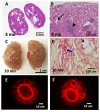
Tns1-KO mice develop features of cystic kidney disease. Tns1-KO kidneys isolated from 3-month-old (A,B) or 10-month-old (C,D) mice were processed for H&E staining (A,B), Sirius Red staining (D), or general morphology (C), showing dilated tubules (black arrows), interstitial infiltration (arrowhead), fibrosis (blue arrows), and cystic kidneys. Representative images of Tns1 WT (E) or KO (F) MDCK in 3D Matrigel culture for 5 days and stained for actin.
Figure 3
Figure 3
Tns2-KO FVB mice develop features of glomerulonephritis. Kidneys isolated from 9-week-old (A) or 6-week-old (B,C) Tns2-KO FBV mice, generated in the laboratory using Tns2-KO embryonic stem cells from the Knockout Mouse Programme (www.mousephenotype.org, accessed on 10 October 2015), were processed and stained with H&E, showing mesangial proliferation (black arrows), segmental glomerulosclerosis (arrowhead), tubular dilatation (asterisk), cast (blue dot), and interstitial infiltration (red arrow). These defects are mouse-strain-dependent and are not developed in the kidneys of Tns2-KO C57BL6 mice, even at 2 years of age (D).
Figure 4
Figure 4
Cumulative survival rate of patients with RCC by tensin status. The Kaplan–Meier curves reveal the cumulative survival rate among patients with CCC (N = 530) and those with PCC (N = 288), stratified according to the status of each tensin. The p values are the result of the log-rank test for the comparison between the two groups. High CTEN (HR = 1.63, p = 0.0023), low TNS1 (HR = 0.38, p < 0.001), and low TNS3 (HR = 0.45, p < 0.001) increase the risk of mortality significantly in patients with CCC. High TNS2 is significantly associated with poor survival in those with PCC (HR = 2, p = 0.026). The association between TNS1, TNS3, and TNS4 and survival in patients with PCC is not significant.

References

    1. Winograd-Katz S.E., Fässler R., Geiger B., Legate K.R. The integrin adhesome: From genes and proteins to human disease. Nat. Rev. Mol. Cell Biol. 2014;15:273–288. doi: 10.1038/nrm3769. - DOI - PubMed
    1. Katz M., Amit I., Citri A., Shay T., Carvalho S., Lavi S., Milanezi F., Lyass L., Amariglio N., Jacob-Hirsch J., et al. A reciprocal tensin-3-cten switch mediates EGF-driven mammary cell migration. Nat. Cell Biol. 2007;9:961–969. doi: 10.1038/ncb1622. - DOI - PubMed
    1. Lo S.H., Janmey P.A., Hartwig J.H., Chen L.B. Interactions of tensin with actin and identification of its three distinct actin-binding domains. J. Cell Biol. 1994;125:1067–1075. doi: 10.1083/jcb.125.5.1067. - DOI - PMC - PubMed
    1. Blangy A. Tensins are versatile regulators of Rho GTPase signalling and cell adhesion. Biol. Cell. 2017;109:115–126. doi: 10.1111/boc.201600053. - DOI - PubMed
    1. Georgiadou M., Ivaska J. Tensins: Bridging AMP-Activated Protein Kinase with Integrin Activation. Trends Cell Biol. 2017;27:703–711. doi: 10.1016/j.tcb.2017.06.004. - DOI - PubMed

LinkOut - more resources