. 2023 May 25;13(6):1253.
doi: 10.3390/life13061253.

Retromode Scanning Laser Ophthalmoscopy for Choroidal Nevi: A Preliminary Study

Affiliations

Retromode Scanning Laser Ophthalmoscopy for Choroidal Nevi: A Preliminary Study

Claudia Azzolini et al. Life (Basel). .

Abstract

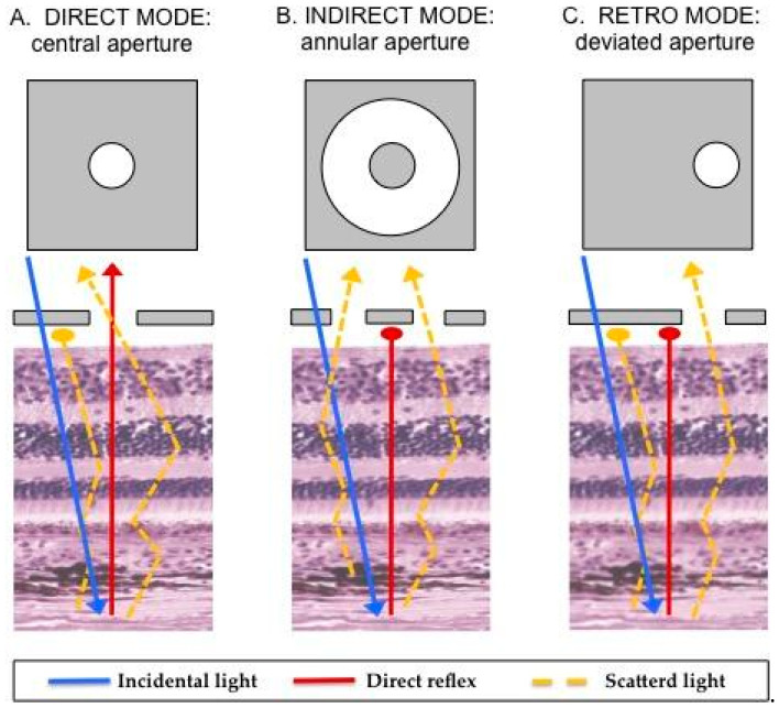
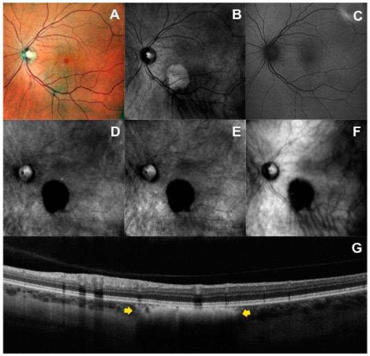
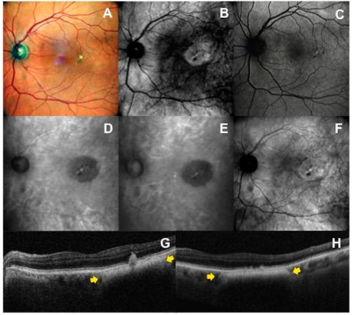
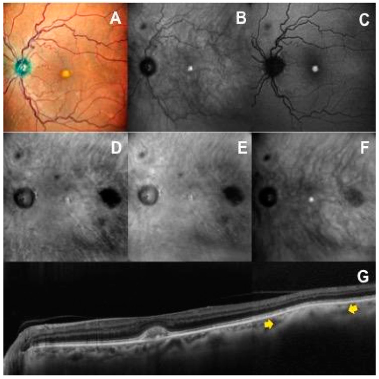
The purpose of the present study was to document pathological findings on retromode imaging in choroidal nevi and evaluate its diagnostic validity, using the confocal scanning laser ophthalmoscope Nidek Mirante (cSLO). A total of 41 choroidal nevi from 41 patients were included. All patients underwent multicolor fundus (mCF), infrared reflectance (IR), green fundus autofluorescence (FAF), dark-field (DF) and retromode (RM) imaging and optical coherence tomography (OCT) scans. We investigated retromode images to evaluate choroidal nevus features by comparing the results with those of mCF, IR, FAF, DF and OCT. In 100% of available images, retromode scanning laser ophthalmoscopy was able to detect choroidal nevi with a characteristic "hypo-retro-reflective" pattern, even the cases not visible on mCF, IR and FAF images. It also made it possible to delineate the margins of lesions with the highest rate of sharpness and accuracy among the imaging modalities examined. These findings seem to demonstrate how RM-SLO is an innovative diagnostic tool to detect and follow up choroidal nevi in a fast, reliable and non-invasive way.

Keywords: choroidal nevus; multimodal imaging; retromode; scanning laser ophthalmoscopy.


Conflict of interest statement

The authors have no conflict of interest to report. This manuscript has not previously been submitted for publication. None of the authors have any financial disclosures to declare.

Figures

Figure 1
Figure 1
Illustration of the mechanism underlying retromode scanning laser ophthalmoscopy [8,9,10,11]. Once the fundus has been illuminated (incidental light), there are two types of light returning back to the instrument detector: a direct reflex and scattered light. Varying the imaging aperture allows the scanning laser ophthalmoscope to assess the light returning from various parts of the eye. (A) Direct confocal mode: a central confocal aperture limits the passage to almost exclusively directly reflected light from the illuminated point on the retina, while other sources of light scatter are blocked. This increases image resolution and contrast. (B) Indirect mode, the so-called “dark-field” mode (RA aperture): a central circular stop blocks the directly reflected light, while more widely scattered light can pass through an annular aperture. This creates low-contrast transillumination images. (C) Retromode (DR or DL aperture): the opening of the ring aperture is restricted and deviates laterally from the confocal light path. The laterally deviated aperture is used to collect the backscattered light from just one direction, and block the directly reflected light and the light scattered from the other directions. This smaller aperture produces higher-contrast transillumination images with a narrower depth of focus compared to the dark-field images.
Figure 2
Figure 2
Multimodal imaging of a patient with choroidal nevus in the left eye: (A) Multicolor fundus image shows a lightly and heterogeneous pigmented area with barely visible lesion borders localized inferior to the fovea. (B) Infrared image shows a hyperreflective mass. (C) On the green fundus autofluorescence image, the nevus is not visualized. Left-deviated (D) and right-deviated (E) retromode images show the characteristic hypo-retro-reflective pattern within the choroidal nevus characterized by a dense dark shadow with sharp margins, shared also with dark-field mode (F). (G) The horizontal optical coherence tomography scan passing through the lesion identifies the choroidal nevus (yellow arrows) as a flat hyperreflective lesion with posterior shadowing, an intact overlying retinal pigment epithelium, and intact outer retinal layers.
Figure 3
Figure 3
Multimodal imaging features in a patient with juxtafoveal choroidal nevus with overlying drusen in the left eye: (A) The nevus is not visible in the multicolor fundus image, except for the presence of multiple yellow drusen-like deposits and pigmentary rearrangement overlying the mass. (B) In the infrared reflectance image, the nevus is well defined as a round hyperreflective area. (C) Fundus autofluorescence imaging cannot image the tumor itself, but shows secondary overlying RPE and retinal changes. Left-deviated (D) and right-deviated (E) retromode images highlight a hypo-retro-reflective mass with a less intense gray shadow and overlying flecks. (F) On dark-field imaging, the choroidal nevus shows an iso-retro-reflective pattern with flecks. (G,H) The corresponding horizontal OCT scans show the choroidal nevus (yellow arrows) and the overlying drusen-like deposits accompanied by changes at the level of the retinal pigment epithelium (e.g., RPE migration, RPE clumping).
Figure 4
Figure 4
A case with a small choroidal nevus in the posterior pole of the left eye not visible on multicolor fundus (A), infrared reflectance (B) and autofluorescence (C) images. The choroidal nevus is clearly detectable on retromode images (D,E) as a dark shadow temporal to the macula, on dark-field image (F) as a gray shadow, and on OCT scan (G) as an hyperreflective lesion with posterior shadowing (yellow arrows).

References

    1. Sumich P., Mitchell P. Choroidal nevi in a white population: The Blue Mountains Eye Study. Arch. Ophthalmol. 1998;116:645–650. doi: 10.1001/archopht.116.5.645. - DOI - PubMed
    1. Chien J.L., Sioufi K. Choroidal nevus: A review of prevalence, features, genetics, risks, and outcomes. Curr. Opin. Ophthalmol. 2017;28:228–237. doi: 10.1097/ICU.0000000000000361. - DOI - PubMed
    1. Singh A.D., Kalyani P. Estimating the risk of malignant transformation of a choroidal nevus. Ophthalmology. 2005;112:1784–1789. doi: 10.1016/j.ophtha.2005.06.011. - DOI - PubMed
    1. Shields C.L., Dalvin L.A. Choroidal nevus imaging features in 3,806 cases and risk factors for transformation into melanoma in 2,355 cases: The 2020 Taylor R. Smith and Victor T. Curtin Lecture. Retina. 2019;39:1840–1851. doi: 10.1097/IAE.0000000000002440. - DOI - PubMed
    1. Geiger F., Said S. Assessing Choroidal Nevi, Melanomas and Indeterminate Melanocytic Lesions Using Multimodal Imaging-A Retrospective Chart Review. Curr. Oncol. 2022;29:1018–1028. doi: 10.3390/curroncol29020087. - DOI - PMC - PubMed