Review
. 2023 May 25;13(6):1254.
doi: 10.3390/life13061254.

Spinal Cord Organoids to Study Motor Neuron Development and Disease

Affiliations
Review

Spinal Cord Organoids to Study Motor Neuron Development and Disease

Felix Buchner et al. Life (Basel). .

Abstract

Motor neuron diseases (MNDs) are a heterogeneous group of disorders that affect the cranial and/or spinal motor neurons (spMNs), spinal sensory neurons and the muscular system. Although they have been investigated for decades, we still lack a comprehensive understanding of the underlying molecular mechanisms; and therefore, efficacious therapies are scarce. Model organisms and relatively simple two-dimensional cell culture systems have been instrumental in our current knowledge of neuromuscular disease pathology; however, in the recent years, human 3D in vitro models have transformed the disease-modeling landscape. While cerebral organoids have been pursued the most, interest in spinal cord organoids (SCOs) is now also increasing. Pluripotent stem cell (PSC)-based protocols to generate SpC-like structures, sometimes including the adjacent mesoderm and derived skeletal muscle, are constantly being refined and applied to study early human neuromuscular development and disease. In this review, we outline the evolution of human PSC-derived models for generating spMN and recapitulating SpC development. We also discuss how these models have been applied to exploring the basis of human neurodevelopmental and neurodegenerative diseases. Finally, we provide an overview of the main challenges to overcome in order to generate more physiologically relevant human SpC models and propose some exciting new perspectives.

Keywords: development; in vitro disease modeling; induced pluripotent stem cells (iPSCs); motor neuron (MN); motor neuron diseases (MNDs); organoids; spinal cord (SpC); spinal cord organoids (SCOs).


Conflict of interest statement

The authors declare no conflict of interest.

Figures

Figure 1
Figure 1
Spinal cord (SpC) organogenesis and recapitulation in vitro. (A) During neurulation—depicted from several Carnegie stages, CS—the neuroectoderm folds and builds a tubular structure that will later develop into the SpC. (B) Inductive signaling from molecular organizer regions, which secrete morphogens, causes the primitive neural tube to be patterned across the DV axis. This patterning is translated into domain-specific expression of transcription factors creating neuronal diversity and heterogeneity. (C) At the same time, the embryo undergoes body column elongation from rostral to caudal, causing the emergence of SpC axial segments. When the neuromesodermal progenitors (NMPs) located in the tail bud are exposed to retinoic acid (RA), released from the adjacent somites, they start to differentiate and adopt colinear HOX expression. (D) In vitro, hiPSCs can be patterned into neural progenitor cells (NPCs)—either in 2D or aggregated in an embryoid body (EB)—through dual SMAD inhibition and may alternatively be induced to differentiate into axial progenitors through WNT-FGF signaling. Created with BioRender.com (accessed on 2 May 2023).
Figure 2
Figure 2
Mimicking the HOX clock to modulate axial elongation in vitro. Most protocols to form spinal cord organoids pattern hiPSCs directly into anterior neural progenitor cells (NPCs), inducing differentiation by RA and SHH pathway activation, and thus, ventral spinal cord fate and formation of hindbrain/cervical spinal motor neurons (spMNs) are achieved. In contrast, the generation of axial progenitors (NMPs) allows for the prolonged activation of the HOX clock. Hereby, the length of exposure to WNT/FGF before RA determines which HOX genes are expressed; therefore, different organoid protocols are designed in a way to give rise to the spMNs of a certain axial identity in a controlled manner. Prolonged maintenance of NMPs in neurobasal media seems to enable the formation of neuromuscular junction organoids, which contain spMNs of mixed axial identity that innervate the skeletal muscle part of the organoid. Created with BioRender.com (accessed on 2 May 2023).
Figure 3
Figure 3
A plethora of options for the protocol optimization of spinal cord organoids (SCOs) can be explored. (A) Through testing the genetic integrity of hiPSC lines, researchers may find cell lines which are optimally suited for organoid modelling studies due to the low prevalence of background mutations. Moreover, disease modeling studies can be improved thoroughly by introducing isogenic lines as alternative controls. (B) Stratification of the SCOs along the dorsoventral (DV) axis may be achieved through protocol optimizations yielding organizer-like regions. (C) Moreover, adding functional vascularization can facilitate nutrient supply and prevent the formation of a necrotic core. (D) Benchmarking the cellular heterogeneity observed in the organoids against scRNAseq datasets from human or murine spinal cords can provide valuable input on which cellular subpopulations are contained in the in vivo organ, and how well the diversity of these cell clusters is recapitulated in an in vitro model. (E) The limited SCO maturation in many protocols can be improved by different small molecule-based strategies. (F) Matrigel, hydrogels and alginate, the most commonly used extracellular matrices for embedding spheroids or organoids. Created with BioRender.com (accessed on 2 May 2023).
Figure 4
Figure 4
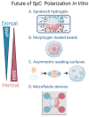
New technological approaches to induce SpC polarization in vitro. (A) Customized “sandwich” hydrogels loaded with BMP and SHH in two separate layers, the organoid is embedded in between both. (B) Organoid grown around beads loaded with SHH. (C) An asymmetrically patterned surface for seeding cells that induces self-organization into dorsal and ventral signaling hubs of the neural tube. (D) A microfluidic device to generate BMP and SHH gradients to dorsalize and ventralize the SCO. Created with BioRender.com (accessed on 2 May 2023).
Figure 5
Figure 5
Overview of applications for spinal cord organoid (SCO) modeling. (A) Studying human spinal cord development by collecting SCOs at different time points allows us to perform longitudinal studies (i.e., scRNAseq analysis, lineage tracing) that can be benchmarked against in vivo counterparts. (B) Assembling multiple organoids or spheroids, fusing them into one coherent structure, allows researchers to study corticomotor circuits. Alternatively, hiPSCs can be induced into neuromesodermal progenitors (NMPs), bipotent cells that can give rise to both neuroectoderm and the cognate skeletal muscle. (C) High-throughput small-molecule screens on SCOs to identify new potential therapeutic targets. (D) SCOs or their derivatives have the potential to become sources for cell transplantation, aiming to ameliorate neuronal loss or restore and regenerate function in neuromuscular diseases. Created with BioRender.com (accessed on 17 May 2023).

References

    1. Goulding M. Circuits controlling vertebrate locomotion: Moving in a new direction. Nat. Rev. Neurosci. 2009;10:507–518. doi: 10.1038/nrn2608. - DOI - PMC - PubMed
    1. Landmesser L.T. The acquisition of motoneuron subtype identity and motor circuit formation. Int. J. Dev. Neurosci. 2001;19:175–182. doi: 10.1016/S0736-5748(00)00090-3. - DOI - PubMed
    1. Stifani N. Motor neurons and the generation of spinal motor neuron diversity. Front. Cell. Neurosci. 2014;8:293. doi: 10.3389/fncel.2014.00293. - DOI - PMC - PubMed
    1. Kadoshima T., Sakaguchi H., Nakano T., Soen M., Ando S., Eiraku M., Sasai Y. Self-organization of axial polarity, inside-out layer pattern, and species-specific progenitor dynamics in human ES cell–derived neocortex. Proc. Natl. Acad. Sci. USA. 2013;110:20284–20289. doi: 10.1073/pnas.1315710110. - DOI - PMC - PubMed
    1. Lancaster M.A., Renner M., Martin C.-A., Wenzel D., Bicknell L.S., Hurles M.E., Homfray T., Penninger J.M., Jackson A.P., Knoblich J.A. Cerebral organoids model human brain development and microcephaly. Nature. 2013;501:373–379. doi: 10.1038/nature12517. - DOI - PMC - PubMed

LinkOut - more resources