Review
. 2023 May 25;13(6):1260.
doi: 10.3390/life13061260.

Mechanisms of Atrial Fibrillation: How Our Knowledge Affects Clinical Practice

Affiliations
Review

Mechanisms of Atrial Fibrillation: How Our Knowledge Affects Clinical Practice

Georgios Leventopoulos et al. Life (Basel). .

Abstract

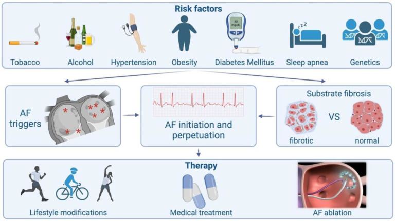
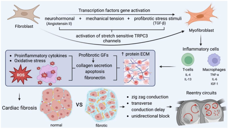
Atrial fibrillation (AF) is a very common arrhythmia that mainly affects older individuals. The mechanism of atrial fibrillation is complex and is related to the pathogenesis of trigger activation and the perpetuation of arrhythmia. The pulmonary veins in the left atrium arei confirm that onfirm the most common triggers due to their distinct anatomical and electrophysiological properties. As a result, their electrical isolation by ablation is the cornerstone of invasive AF treatment. Multiple factors and comorbidities affect the atrial tissue and lead to myocardial stretch. Several neurohormonal and structural changes occur, leading to inflammation and oxidative stress and, consequently, a fibrotic substrate created by myofibroblasts, which encourages AF perpetuation. Several mechanisms are implemented into daily clinical practice in both interventions in and the medical treatment of atrial fibrillation.

Keywords: atrial fibrillation; fibrosis; inflammation oxidative stress; triggers.


Conflict of interest statement

The authors declare no conflict of interest.

Figures

Figure 1
Figure 1
Schematic presentation of atrial fibrillation from causes to treatment.
Figure 2
Figure 2
(Left) Mechanism that explains how tissue stretch promotes atrial premature beats, mainly through afterdepolarization. (Right) Specific electrophysiologic properties of the pulmonary veins render them more likely to be the origin of atrial ectopic beats. NADPH oxidase (nicotinamide adenine dinucleotide phosphate oxidase) EAD: early afterdepolarization. DAD: delayed afterdepolarization. RMP: resting membrane potential.
Figure 3
Figure 3
A. Atrial ectopic beat originates from the proximal aspect of the coronary sinus (arrow at the CS 7-8 electrogram)—a non PV trigger—and induces atrial fibrillation (G. Leventopoulos’ archive). PV: pulmonary vein. CS: coronary sinus.
Figure 4
Figure 4
(Bottom left) Left atrium—posterior view. All 4 veins and the posterior wall are illustrated; they share the same embryologic origin. (Top right) Left atrium—anterior view. Note the left atrial appendage and the coronary sinus (catheter placed inside), which are rarely additional PV triggers. (Bottom right) Left anterior oblique view. The anatomical correlation between right and left atrium is depicted. Geometry of both atria was created in a patient with perimitral flutter, in this particular case. Generally, several additional PV triggers are also located in the right atrium, such as the superior vena cava, the right atrial appendage, the crista terminalis, and the tricuspid valve. Occasionally, AF involves the degeneration of AVNRT (atrioventricular nodal reciprocating tachycardia). * Potential anatomical origins of atrial ectopic beat.
Figure 5
Figure 5
The key role of myofibroblasts in fibrosis formation and the main pathophysiologic cascades. Mechanical stress accounts for myocardial injury, oxidative stress, and neurohormonal activation, which transform fibroblasts into myofibroblasts, which, along with other inflammatory cells, encourage fibrotic substrates. In this microenviroment, all the required conditions for the formation and perpetuation of reentry circuits are fulfilled and further enhanced. GF: growth factor, IL: interleukin, TGF: transforming growth factor, TNF, tumor necrosis factor. IGF: insulin growth factor.

References

    1. Heeringa J., van der Kuip D.A., Hofman A., Kors J.A., van Herpen G., Stricker BH C., Stijnen T., Lip G.Y.H., Witteman J.C.M. Prevalence, incidence and lifetime risk of atrial fibrillation: The Rotterdam study. Eur. Heart J. 2006;27:949–953. doi: 10.1093/eurheartj/ehi825. - DOI - PubMed
    1. Zoni-Berisso M., Lercari F., Carazza T., Domenicucci S. Epidemiology of atrial fibrillation: European perspective. Clin. Epidemiol. 2014;6:213–220. doi: 10.2147/CLEP.S47385. - DOI - PMC - PubMed
    1. Kornej J., Börschel C.S., Benjamin E.J., Schnabel R.B. Epidemiology of Atrial Fibrillation in the 21st CenturyNovel Methods and New Insights. Circ. Res. 2020;127:4–20. doi: 10.1161/CIRCRESAHA.120.316340. - DOI - PMC - PubMed
    1. Tsigkas G., Apostolos A., Despotopoulos S., Vasilagkos G., Kallergis E., Leventopoulos G., Mplani V., Davlouros P. Heart failure and atrial fibrillation: New concepts in pathophysiology, management, and future directions. Heart Fail. Rev. 2022;27:1201–1210. doi: 10.1007/s10741-021-10133-6. - DOI - PubMed
    1. Olshansky B., Goldberger Z.D., FACC. FHRS. Pogwizd S.M. The electrocardiogram in atrial. In: Knight B.P., Yeon S.B., editors. UpToDate. UpToDate; Waltham, MA, USA: 2021.