A Comparative Study of the Synthesis and Characterization of Biogenic Selenium Nanoparticles by Two Contrasting Endophytic Selenobacteria
- PMID: 37375102
- PMCID: PMC10303461
- DOI: 10.3390/microorganisms11061600
A Comparative Study of the Synthesis and Characterization of Biogenic Selenium Nanoparticles by Two Contrasting Endophytic Selenobacteria
Abstract
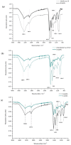
The present study examined the biosynthesis and characterization of selenium nanoparticles (SeNPs) using two contrasting endophytic selenobacteria, one Gram-positive (Bacillus sp. E5 identified as Bacillus paranthracis) and one Gram-negative (Enterobacter sp. EC5.2 identified as Enterobacter ludwigi), for further use as biofortifying agents and/or for other biotechnological purposes. We demonstrated that, upon regulating culture conditions and selenite exposure time, both strains were suitable "cell factories" for producing SeNPs (B-SeNPs from B. paranthracis and E-SeNPs from E. ludwigii) with different properties. Briefly, dynamic light scattering (DLS), transmission electron microscopy (TEM), and atomic force microscopy (AFM) studies revealed that intracellular E-SeNPs (56.23 ± 4.85 nm) were smaller in diameter than B-SeNPs (83.44 ± 2.90 nm) and that both formulations were located in the surrounding medium or bound to the cell wall. AFM images indicated the absence of relevant variations in bacterial volume and shape and revealed the existence of layers of peptidoglycan surrounding the bacterial cell wall under the conditions of biosynthesis, particularly in the case of B. paranthracis. Raman spectroscopy, Fourier transform infrared spectroscopy (FTIR), energy-dispersive X-ray (EDS), X-ray diffraction (XRD), and X-ray photoelectron spectroscopy (XPS) showed that SeNPs were surrounded by the proteins, lipids, and polysaccharides of bacterial cells and that the numbers of the functional groups present in B-SeNPs were higher than in E-SeNPs. Thus, considering that these findings support the suitability of these two endophytic stains as potential biocatalysts to produce high-quality Se-based nanoparticles, our future efforts must be focused on the evaluation of their bioactivity, as well as on the determination of how the different features of each SeNP modulate their biological action and their stability.
Keywords: Bacillus paranthracis; Enterobacter ludwigii; biofortification; biogenic nanoparticles; selenite; selenium.
Conflict of interest statement
The authors declare that they have no known competing financial interests or personal relationships that could have appeared to influence the work reported in this paper.
Figures


Similar articles
-
A newly isolated Bacillus amyloliquefaciens SRB04 for the synthesis of selenium nanoparticles with potential antibacterial properties.Int Microbiol. 2021 Jan;24(1):103-114. doi: 10.1007/s10123-020-00147-9. Epub 2020 Oct 29. Int Microbiol. 2021. PMID: 33124680
-
Preparation, characteristics and antioxidant activity of polysaccharides and proteins-capped selenium nanoparticles synthesized by Lactobacillus casei ATCC 393.Carbohydr Polym. 2018 Sep 1;195:576-585. doi: 10.1016/j.carbpol.2018.04.110. Epub 2018 Apr 30. Carbohydr Polym. 2018. PMID: 29805014
-
Nitrate reductase involves in selenite reduction in Rahnella aquatilis HX2 and the characterization and anticancer activity of the biogenic selenium nanoparticles.J Trace Elem Med Biol. 2024 May;83:127387. doi: 10.1016/j.jtemb.2024.127387. Epub 2024 Jan 11. J Trace Elem Med Biol. 2024. PMID: 38237425
-
Selenite Reduction by Proteus sp. YS02: New Insights Revealed by Comparative Transcriptomics and Antibacterial Effectiveness of the Biogenic Se0 Nanoparticles.Front Microbiol. 2022 Mar 10;13:845321. doi: 10.3389/fmicb.2022.845321. eCollection 2022. Front Microbiol. 2022. PMID: 35359742 Free PMC article.
-
Physical-Chemical Properties of Biogenic Selenium Nanostructures Produced by Stenotrophomonas maltophilia SeITE02 and Ochrobactrum sp. MPV1.Front Microbiol. 2018 Dec 19;9:3178. doi: 10.3389/fmicb.2018.03178. eCollection 2018. Front Microbiol. 2018. PMID: 30619230 Free PMC article.
Cited by
-
Harnessing selenium nanoparticles (SeNPs) for enhancing growth and germination, and mitigating oxidative stress in Pisum sativum L.Sci Rep. 2023 Nov 21;13(1):20379. doi: 10.1038/s41598-023-47616-5. Sci Rep. 2023. PMID: 37989844 Free PMC article.
-
Selenium nanoparticle loaded on PVA/chitosan biofilm synthesized from orange peels: antimicrobial and antioxidant properties for plum preservation.BMC Chem. 2025 Aug 20;19(1):245. doi: 10.1186/s13065-025-01608-w. BMC Chem. 2025. PMID: 40835945 Free PMC article.
-
Antioxidant and Anticancer Activities of Bio-Synthesized Selenium Nanoparticles by Escherichia coli.Asian Pac J Cancer Prev. 2025 Apr 1;26(4):1303-1311. doi: 10.31557/APJCP.2025.26.4.1303. Asian Pac J Cancer Prev. 2025. PMID: 40302083 Free PMC article.
-
Colistin-Conjugated Selenium Nanoparticles: A Dual-Action Strategy Against Drug-Resistant Infections and Cancer.Pharmaceutics. 2025 Apr 24;17(5):556. doi: 10.3390/pharmaceutics17050556. Pharmaceutics. 2025. PMID: 40430850 Free PMC article.
References
-
- El-Ramady H.R., Domokos-Szabolcsy É., Abdalla N.A., Alshaal T.A., Shalaby T.A., Sztrik A., Prokisch J., Fári M. Selenium and nano-selenium in agroecosystems. Environ. Chem. Lett. 2014;12:495–510. doi: 10.1007/s10311-014-0476-0. - DOI
-
- Kabata-Pendias A. Trace Elements in Soils and Plants. 4th ed. CRC Press; Boca Raton, FL, USA: 2010.
LinkOut - more resources
Full Text Sources
Miscellaneous

