Review
. 2023 Jun 19;11(6):1614.
doi: 10.3390/microorganisms11061614.

Microbial Biofilm: A Review on Formation, Infection, Antibiotic Resistance, Control Measures, and Innovative Treatment

Affiliations
Review

Microbial Biofilm: A Review on Formation, Infection, Antibiotic Resistance, Control Measures, and Innovative Treatment

Satish Sharma et al. Microorganisms. .

Erratum in

Abstract

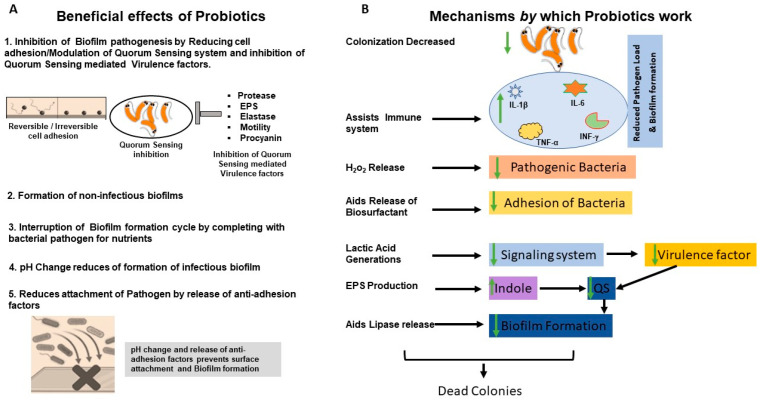
Biofilm is complex and consists of bacterial colonies that reside in an exopolysaccharide matrix that attaches to foreign surfaces in a living organism. Biofilm frequently leads to nosocomial, chronic infections in clinical settings. Since the bacteria in the biofilm have developed antibiotic resistance, using antibiotics alone to treat infections brought on by biofilm is ineffective. This review provides a succinct summary of the theories behind the composition of, formation of, and drug-resistant infections attributed to biofilm and cutting-edge curative approaches to counteract and treat biofilm. The high frequency of medical device-induced infections due to biofilm warrants the application of innovative technologies to manage the complexities presented by biofilm.

Keywords: antibiotic resistance; biofilm; biofilm control; extracellular polysaccharides; healthcare-associated infection; medical device infections.


Conflict of interest statement

The authors declare no conflict of interest.

Figures

Figure 1
Figure 1
A conventional scanning electron microscopic (SEM) image of a biofilm formed by M. haemolytica (D153), grown in colorless RPMI 1640 (AC) and S. aureus Newbould 305 (NB305), grown in BHI broth (DF) on round glass coverslips in 24-well plates at 37 °C. Biofilms grown on glass coverslips were fixed with 10% formalin (A,D), 2.5% glutaraldehyde (B,E), or Methacarn (C,F) fixative solutions for 48 h and samples were further processed for SEM examination. EPS layers on the top, in the middle, and in the bottom of biofilms (C,F) are shown by white arrows. This figure is reproduced from PLoS One. 2020; 15(5): e0233973. This is an open access article, free of all copyright, and is reproduced under the Creative Commons CC0 public domain dedication.
Figure 2
Figure 2
Diagrammatic illustration showing the growth cycle of a biofilm by a single bacterium species on a solid surface. (1) Reversible attachment of single planktonic bacteria to surfaces. The first attachment of the bacteria is influenced by attractive or repelling forces generated by nutrient levels, pH, and the temperature of the surface. (2) Aggregation of bacteria and irreversible attachment to surfaces. (3) Formation of an external matrix of multilayered complex biomolecules, microcolony formation, and EPS secretion that constitute the external matrix. Secretion of polysaccharides in biofilm forming strains enables aggregation, adherence, and surface tolerance, allowing for improved surface colonization. (4) Maturation of biofilms and acquisition of a three-dimensional structure as they reach maturity. These three-dimensional structures rest on self-produced extracellular matrix components. (5) Fully mature biofilms detach, which allows bacterial cells to take on a planktonic state once again and thereby establish biofilm in other locations. Created on BioRender.com 31 March 2023.
Figure 3
Figure 3
Biofilm formation and pathogenesis mechanism of CAUTI: The environmental conditions created on the catheter surface make it an ideal site for bacterial attachment and formation of biofilm structures. (1) Bacteria migrates through the periurethral area along the catheter surface. (2) Fimbriae attach to the body-fluid-derived catheter surface or directly to the catheter material inducing EPS production and biofilm formation. (3) Some bacteria such as P. mirabilis produce enzymes involved in the hydrolysis of urea in urine into ammonia, increasing the local pH leading to the production of minerals in urine which results in struvite crystals. (4) Struvite formed is incorporated into the developing biofilm—a process called ureolytic mineralization, which is also facilitated by the capsule polysaccharides. (5) Fully developed crystalline biofilm eventually causes catheter obstruction. Created on Biorender.com (31 March 2023).
Figure 4
Figure 4
Conventional and novel methods to control biofilm: To fight infections ensuing from biofilms, numerous methods have been developed from different aspects; some are conventional, and some are novel. (1) Use of conventional antibiotics in the early stage; however, this method has a high failure rate due to poor penetration and lack of action due to hypoxia. (2) Phage therapy has been used as an alternate approach for controlling biofilm formation. This method works by depolarizing the EPS to disrupt the biofilm. This method also has multiple limitations including resistance, clearance by the host immune system, and specific only to certain strains of bacteria. (3) Novel methods of biofilm disruption such as QS system inhibitors which interfere with the microbial communication mechanism based on molecular signatures are also being used. (4) Newer methods such as antibody-based therapy against biofilm are being tried in preclinical models that target several biofilms but are limited in their success due to poor target specificity and infusion reaction. (5) Natural product-based therapy is another conventional method used. Products used here are either crude extract or purified compounds. Biologically active compounds showing antibacterial activity are extracted, purified, and successfully evaluated to clinical and pre-clinical models. Such extracts include chloroform, ethanol, and methyl ester. These extracts inhibit biofilm through various mechanisms ranging from inhibition of a critical enzyme involved in the growth of bacteria to repression of gene expression of multiple genes required for bacterial growth and development.
Figure 5
Figure 5
Probiotic methods to control biofilms. Probiotics pathogenic biofilm inhibition in different ways, as shown in the Figure. (A) Beneficial effects of probiotics (B) Mechanism of action of probiotics.

References

    1. Bjarnsholt T., Ciofu O., Molin S., Givskov M., Hoiby N. Applying insights from biofilm biology to drug development—Can a new approach be developed? Nat. Rev. Drug. Discov. 2013;12:791–808. doi: 10.1038/nrd4000. - DOI - PubMed
    1. Irie Y., Borlee B.R., O’Connor J.R., Hill P.J., Harwood C.S., Wozniak D.J., Parsek M.R. Self-produced exopolysaccharide is a signal that stimulates biofilm formation in Pseudomonas aeruginosa. Proc. Natl. Acad. Sci. USA. 2012;109:20632–20636. doi: 10.1073/pnas.1217993109. - DOI - PMC - PubMed
    1. Gupta P., Sarkar S., Das B., Bhattacharjee S., Tribedi P. Biofilm, pathogenesis and prevention--a journey to break the wall: A review. Arch. Microbiol. 2016;198:1–15. doi: 10.1007/s00203-015-1148-6. - DOI - PubMed
    1. Costerton J.W., Geesey G.G., Cheng K.J. How bacteria stick. Sci. Am. 1978;238:86–95. doi: 10.1038/scientificamerican0178-86. - DOI - PubMed
    1. Costerton J.W., Stewart P.S., Greenberg E.P. Bacterial biofilms: A common cause of persistent infections. Science. 1999;284:1318–1322. doi: 10.1126/science.284.5418.1318. - DOI - PubMed

LinkOut - more resources