. 2023 Jun 15;28(12):4777.
doi: 10.3390/molecules28124777.

Synthesis, Characterization, DFT Studies of Novel Cu(II), Zn(II), VO(II), Cr(III), and La(III) Chloro-Substituted Schiff Base Complexes: Aspects of Its Antimicrobial, Antioxidant, Anti-Inflammatory, and Photodegradation of Methylene Blue

Affiliations

Synthesis, Characterization, DFT Studies of Novel Cu(II), Zn(II), VO(II), Cr(III), and La(III) Chloro-Substituted Schiff Base Complexes: Aspects of Its Antimicrobial, Antioxidant, Anti-Inflammatory, and Photodegradation of Methylene Blue

Laila H Abdel-Rahman et al. Molecules. .

Abstract

A new chlorobenzylidene imine ligand, (E)-1-((5-chloro-2-hydroxybenzylidene)amino) naphthalen-2-ol (HL), and its [Zn(L)(NO3)(H2O)3], [La(L)(NO3)2(H2O)2], [VO(L)(OC2H5)(H2O)2], [Cu(L)(NO3)(H2O)3], and [Cr(L)(NO3)2(H2O)2], complexes were synthesized and characterized. The characterization involved elemental analysis, FT-IR, UV/Vis, NMR, mass spectra, molar conductance, and magnetic susceptibility measurements. The obtained data confirmed the octahedral geometrical structures of all metal complexes, while the [VO(L)(OC2H5)(H2O)2] complex exhibited a distorted square pyramidal structure. The complexes were found to be thermally stable based on their kinetic parameters determined using the Coats-Redfern method. The DFT/B3LYP technique was employed to calculate the optimized structures, energy gaps, and other important theoretical descriptors of the complexes. In vitro antibacterial assays were conducted to evaluate the complexes' potential against pathogenic bacteria and fungi, comparing them to the free ligand. The compounds exhibited excellent fungicidal activity against Candida albicans ATCC: 10231 (C. albicans) and Aspergillus negar ATCC: 16404 (A. negar), with inhibition zones of HL, [Zn(L)(NO3)(H2O)3], and [La(L)(NO3)2(H2O)2] three times higher than that of the Nystatin antibiotic. The DNA binding affinity of the metal complexes and their ligand was investigated using UV-visible, viscosity, and gel electrophoresis methods, suggesting an intercalative binding mode. The absorption studies yielded Kb values ranging from 4.40 × 10⁵ to 7.30 × 10⁵ M-1, indicating high binding strength to DNA comparable to ethidium bromide (value 107 M-1). Additionally, the antioxidant activity of all complexes was measured and compared to vitamin C. The anti-inflammatory efficacy of the ligand and its metal complexes was evaluated, revealing that [Cu(L)(NO3)(H2O)3] exhibited the most effective activity compared to ibuprofen. Molecular docking studies were conducted to explore the binding nature and affinity of the synthesized compounds with the receptor of Candida albicans oxidoreductase/oxidoreductase INHIBITOR (PDB ID: 5V5Z). Overall, the combined findings of this work demonstrate the potential of these new compounds as efficient fungicidal and anti-inflammatory agents. Furthermore, the photocatalytic effect of the Cu(II) Schiff base complex/GO was examined.

Keywords: DFT; DNA-binding; Schiff bases; anti-inflammatory; antimicrobial; antioxidant; photodegradation.


Conflict of interest statement

The authors declare no conflict of interest.

Figures
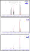
Figure 1
Figure 1
1HNMR spectra of HL ligand (a) and its [Zn(L)(NO3)(H2O)3] (b) and [La(L)(NO3)2(H2O)2] (c) complexes.
Scheme 1
Scheme 1
Mass fragmentation pattern of the HL Schiff base ligand.
Scheme 2
Scheme 2
Schematic representation for the synthesis of HL Schiff base ligand and its metal complexes.
Figure 2
Figure 2
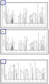
EI-mass spectral analysis of (a) [Cu(L)(NO3)(H2O)3], (b) [Zn(L)(NO3)(H2O)3], (c) [La(L)(NO3)2(H2O)2], and (d) [VO(L)(OC2H5)(H2O)2] complexes.
Figure 2
Figure 2
EI-mass spectral analysis of (a) [Cu(L)(NO3)(H2O)3], (b) [Zn(L)(NO3)(H2O)3], (c) [La(L)(NO3)2(H2O)2], and (d) [VO(L)(OC2H5)(H2O)2] complexes.
Figure 3
Figure 3
pH stability profile of the [Cu(L)(NO3)(H2O)3], [Cr(L)(NO3)2(H2O)2], [La(L)(NO3)2(H2O)2], [VO(L)(OC2H5)(H2O)2], and [Zn(L)(NO3)(H2O)3], complexes in DMF at different pH values.
Figure 4
Figure 4
Natural charges on atoms, the molecular electrostatic potential (MEP) surface by density function B3LYP/6-311++g(d, p), the vector of the dipole moment, and the optimal ligand structure.
Figure 5
Figure 5
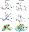
The optimized structure, the vector of the dipole moment, the natural charges on atoms, and the molecular electrostatic potential (MEP) surface on active centers of [CuLNO3(H2O)3] and [ZnLNO3(H2O)3] complexes.
Figure 5
Figure 5
The optimized structure, the vector of the dipole moment, the natural charges on atoms, and the molecular electrostatic potential (MEP) surface on active centers of [CuLNO3(H2O)3] and [ZnLNO3(H2O)3] complexes.
Figure 6
Figure 6
The molecular electrostatic potential (MEP) surface on active centers of the optimized structure of [CrL(NO3)2(H2O)2] and [LaL(NO3)2(H2O)2] complexes, the dipole moment’s vector, the atoms’ natural charges.
Figure 7
Figure 7
The optimized structure, the vector of the dipole moment, the natural charges on atoms, and the molecular electrostatic potential (MEP) surface on active centers of [VOL(H2O)OEt] complex.
Figure 8
Figure 8
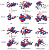
HOMO and LUMO charge density maps of HL, [CuLNO3(H2O)3], [ZnLNO3(H2O)3], [CrL(NO3)2(H2O)2], [LaL(NO3)2(H2O)2], and [VOL(H2O)OEt].
Figure 9
Figure 9
(a) Graphical representation of antimicrobial activity results of HL ligand and its new [Cu(L)(NO3)(H2O)3], [Zn(L)(NO3)(H2O)3], [VO(L)(OC2H5)(H2O)2], [Cr(L)(NO3)2(H2O)2], and [La(L)(NO3)2(H2O)2] complexes at 20 µg/mL concentration and (b) zone of inhibition of [Zn(L)(NO3)(H2O)3], [La(L)(NO3)2(H2O)2], and [VO(L)(OC2H5)(H2O)2] against C. albicans.
Figure 10
Figure 10
Absorption spectra of the complexes [VO(L)(OC2H5)(H2O)2], [Cu(L)(NO3)(H2O)3], [La(L)(NO3)2(H2O)2], [Cr(L)(NO3)2(H2O)2], [Zn(L)(NO3)(H2O)3], and HL in the presence of increasing concentrations of CT-DNA (in Tris-HCl/NaCl buffer). With increasing concentrations of CT-DNA, the absorbance of the complex changes at [complex] = 10 μM and [DNA] = 10–100 μM.
Figure 10
Figure 10
Absorption spectra of the complexes [VO(L)(OC2H5)(H2O)2], [Cu(L)(NO3)(H2O)3], [La(L)(NO3)2(H2O)2], [Cr(L)(NO3)2(H2O)2], [Zn(L)(NO3)(H2O)3], and HL in the presence of increasing concentrations of CT-DNA (in Tris-HCl/NaCl buffer). With increasing concentrations of CT-DNA, the absorbance of the complex changes at [complex] = 10 μM and [DNA] = 10–100 μM.
Figure 11
Figure 11
The relative viscosity of CT-DNA is affected by the quantity of ethidium bromide (EB) and the metal complexes.
Figure 12
Figure 12
Gel electrophoresis pattern showing the interactions of the new complexes with DNA based on gel electrophoresis. Lane 1: DNA Ladder, Lane 2: HL + DNA, Lane 3: [Cu(L)(NO3)(H2O)3]  +  DNA, Lane 4: [VO(L)(OC2H5)(H2O)2] +  DNA, Lane 5: [La(L)(NO3)2(H2O)2] + DNA, and Lane 6: [Zn(L)(NO3)(H2O)3]  +  DNA, Lane 7: [CrL(NO3)2(H2O)2] + DNA.
Figure 13
Figure 13
Concentration-dependent IC50 values of metal complexes’ DPPH radical-scavenging activity.
Figure 14
Figure 14
Inhibition % of protein denaturation of HL ligand and its metal complexes compared to ibuprofen at different concentrations.
Figure 15
Figure 15
Two-dimensional and three-dimensional plots of the interactions between HL, [CuLNO3(H2O)3], and [ZnLNO3(H2O)3] with the active site of the receptor of Candida albicans (PDB ID: PDB ID: 5V5Z). Hydrophobic interactions with amino acid residues are shown with dotted curves.
Figure 16
Figure 16
Two-dimensional and three-dimensional plots of the interactions between [CrL(NO3)2(H2O)2], [LaL(NO3)2(H2O)2], and [VOL(H2O)OEt] with the active site of the receptor of Candida albicans (PDB ID: PDB ID: 5V5Z). Hydrophobic interactions with amino acid residues are shown with dotted curves.
Figure 16
Figure 16
Two-dimensional and three-dimensional plots of the interactions between [CrL(NO3)2(H2O)2], [LaL(NO3)2(H2O)2], and [VOL(H2O)OEt] with the active site of the receptor of Candida albicans (PDB ID: PDB ID: 5V5Z). Hydrophobic interactions with amino acid residues are shown with dotted curves.
Figure 17
Figure 17
(a) The effect of Cu(II) Schiff base complex on MB photocatalytic degradation, [complex] = 2.5 mg, 30 mL of MB. (b) The temporal absorption spectrum changes of MB taking place under visible light irradiation for CuMGO catalyst, initial concentration of MB: 6.25 × 10⁻³ M, 30 mL, CuMGO: 2.5 mg and pH: 10. (c) Optimization of the pH for the degradation of MB. (d) Optimization of the amount of catalyst for degradation and (b) the recyclability of the catalyst.
Figure 17
Figure 17
(a) The effect of Cu(II) Schiff base complex on MB photocatalytic degradation, [complex] = 2.5 mg, 30 mL of MB. (b) The temporal absorption spectrum changes of MB taking place under visible light irradiation for CuMGO catalyst, initial concentration of MB: 6.25 × 10⁻³ M, 30 mL, CuMGO: 2.5 mg and pH: 10. (c) Optimization of the pH for the degradation of MB. (d) Optimization of the amount of catalyst for degradation and (b) the recyclability of the catalyst.
Figure 18
Figure 18
The possible photocatalytic mechanism for the degradation of MB in the presence of Cu(II) Schiff base complex loaded on graphene oxide and H2O2.

References

    1. Abd El-Halim H.F.A., Mohamed G.G., Khalil E.A. Synthesis, spectral, thermal and biological studies of mixed ligand complexes with newly prepared Schiff base and 1,10-phenanthroline ligands. J. Mol. Struct. 2017;1146:153–163. doi: 10.1016/j.molstruc.2017.05.092. - DOI
    1. Mohamed R.G., Elantabli F.M., Helal N.H., El-Medani S.M. New group 6 metal carbonyl complexes with 4,5-dimethyl-N,N-bis(pyridine-2-yl-methylene)benzene-1,2-diimine Schiff base: Synthesis, spectral, cyclic voltammetry and biological activity studies. Spectrochim. Acta Part A Mol. Biomol. Spectrosc. 2015;141:316–326. doi: 10.1016/j.saa.2015.01.054. - DOI - PubMed
    1. Ali O.A.M. Palladium(II) and zinc(II) complexes of neutral [N2O2] donor Schiff bases derived from furfuraldehyde: Synthesis, characterization, fluorescence and corrosion inhibitors of ligands. Spectrochim. Acta Part A Mol. Biomol. Spectrosc. 2014;132:52–60. doi: 10.1016/j.saa.2014.03.127. - DOI - PubMed
    1. Nasrin D., Alam M.A., Hossain M.N., Nazimuddin M. Synthesis, Characterization and Antimicrobial Activity of Metal Complexes of Schiff ’ s Base Derived from S-benzyldithiocarbazate with 2-hydroxyacetophenone. Chem. J. 2013;3:13–19.
    1. Sherif O.E., Abdel-Kader N.S. Spectroscopic and biological activities studies of bivalent transition metal complexes of Schiff bases derived from condensation of 1,4-phenylenediamine and benzopyrone derivatives. Spectrochim. Acta Part A Mol. Biomol. Spectrosc. 2014;117:519–526. doi: 10.1016/j.saa.2013.08.037. - DOI - PubMed

LinkOut - more resources