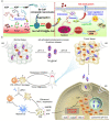
Calcium Phosphate-Based Nanomaterials: Preparation, Multifunction, and Application for Bone Tissue Engineering
- PMID: 37375345
- PMCID: PMC10302440
- DOI: 10.3390/molecules28124790
Calcium Phosphate-Based Nanomaterials: Preparation, Multifunction, and Application for Bone Tissue Engineering
Abstract
Calcium phosphate is the main inorganic component of bone. Calcium phosphate-based biomaterials have demonstrated great potential in bone tissue engineering due to their superior biocompatibility, pH-responsive degradability, excellent osteoinductivity, and similar components to bone. Calcium phosphate nanomaterials have gained more and more attention for their enhanced bioactivity and better integration with host tissues. Additionally, they can also be easily functionalized with metal ions, bioactive molecules/proteins, as well as therapeutic drugs; thus, calcium phosphate-based biomaterials have been widely used in many other fields, such as drug delivery, cancer therapy, and as nanoprobes in bioimaging. Thus, the preparation methods of calcium phosphate nanomaterials were systematically reviewed, and the multifunction strategies of calcium phosphate-based biomaterials have also been comprehensively summarized. Finally, the applications and perspectives of functionalized calcium phosphate biomaterials in bone tissue engineering, including bone defect repair, bone regeneration, and drug delivery, were illustrated and discussed by presenting typical examples.
Keywords: bioimaging; bone tissue engineering; calcium phosphate; drug delivery; multifunction; nanomaterials.
Conflict of interest statement
The authors declare no conflict of interest.
Figures


Similar articles
-
Nanostructured Calcium-based Biomaterials and their Application in Drug Delivery.Curr Med Chem. 2020;27(31):5189-5212. doi: 10.2174/0929867326666190222193357. Curr Med Chem. 2020. PMID: 30806303 Review.
-
[Research development and prospect of calcium phosphate biomaterials with intrinsic osteoinductivity].Sheng Wu Yi Xue Gong Cheng Xue Za Zhi. 2006 Apr;23(2):442-5, 454. Sheng Wu Yi Xue Gong Cheng Xue Za Zhi. 2006. PMID: 16706385 Review. Chinese.
-
Design strategies and applications of nacre-based biomaterials.Acta Biomater. 2017 May;54:21-34. doi: 10.1016/j.actbio.2017.03.003. Epub 2017 Mar 6. Acta Biomater. 2017. PMID: 28274766 Review.
-
Calcium and phosphate ions as simple signaling molecules with versatile osteoinductivity.Biomed Mater. 2018 Jun 14;13(5):055005. doi: 10.1088/1748-605X/aac7a5. Biomed Mater. 2018. PMID: 29794341
-
Recent progress in preparation and applications of chitosan/calcium phosphate composite materials.Int J Biol Macromol. 2021 May 1;178:240-252. doi: 10.1016/j.ijbiomac.2021.02.143. Epub 2021 Feb 22. Int J Biol Macromol. 2021. PMID: 33631262 Review.
Cited by
-
Biomaterials for Drug Delivery and Human Applications.Materials (Basel). 2024 Jan 18;17(2):456. doi: 10.3390/ma17020456. Materials (Basel). 2024. PMID: 38255624 Free PMC article. Review.
-
Designing Dual-Responsive Drug Delivery Systems: The Role of Phase Change Materials and Metal-Organic Frameworks.Materials (Basel). 2024 Jun 22;17(13):3070. doi: 10.3390/ma17133070. Materials (Basel). 2024. PMID: 38998154 Free PMC article. Review.
-
Investigating the association between calcium-phosphorus balance and osteoarthritis: Evidence from NHANES 2007-2016.Medicine (Baltimore). 2025 Jul 18;104(29):e43301. doi: 10.1097/MD.0000000000043301. Medicine (Baltimore). 2025. PMID: 40696700 Free PMC article.
-
Biomimetic Scaffolds of Calcium-Based Materials for Bone Regeneration.Biomimetics (Basel). 2024 Aug 24;9(9):511. doi: 10.3390/biomimetics9090511. Biomimetics (Basel). 2024. PMID: 39329533 Free PMC article. Review.
-
Mechanical Properties and Liquid Absorption of Calcium Phosphate Composite Cements.Materials (Basel). 2023 Aug 17;16(16):5653. doi: 10.3390/ma16165653. Materials (Basel). 2023. PMID: 37629944 Free PMC article.
References
Publication types
MeSH terms
Substances
Grants and funding
LinkOut - more resources
Full Text Sources

