. 2023 Jun 17;28(12):4826.
doi: 10.3390/molecules28124826.

The Phthalic Selenoanhydride Decreases Rat Blood Pressure and Tension of Isolated Mesenteric, Femoral and Renal Arteries

Affiliations

The Phthalic Selenoanhydride Decreases Rat Blood Pressure and Tension of Isolated Mesenteric, Femoral and Renal Arteries

Peter Balis et al. Molecules. .

Abstract

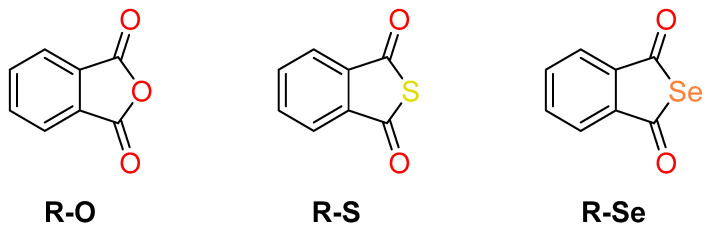
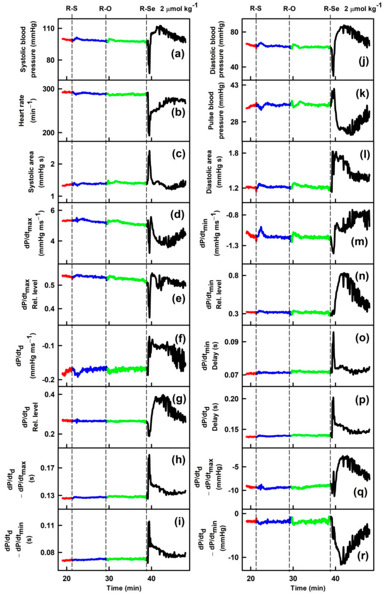
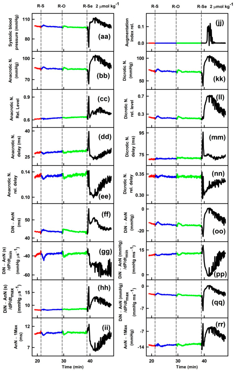
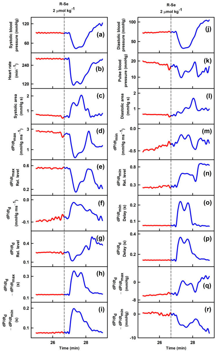
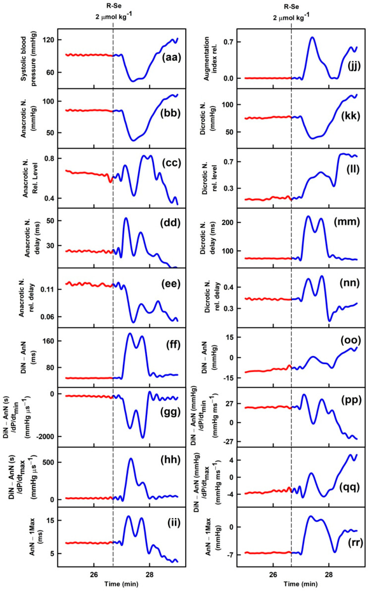
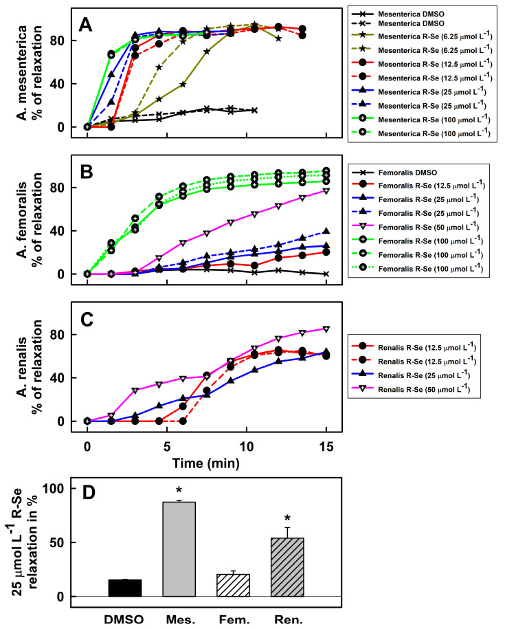
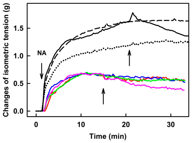
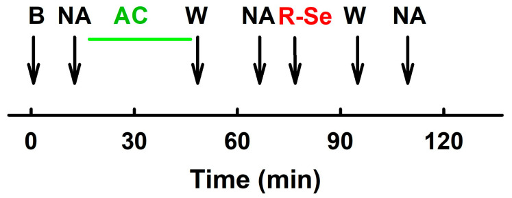
Phthalic selenoanhydride (R-Se) solved in physiological buffer releases various reactive selenium species including H2Se. It is a potential compound for Se supplementation which exerts several biological effects, but its effect on the cardiovascular system is still unknown. Therefore, herein we aimed to study how R-Se affects rat hemodynamic parameters and vasoactive properties in isolated arteries. The right jugular vein of anesthetized Wistar male rats was cannulated for IV administration of R-Se. The arterial pulse waveform (APW) was detected by cannulation of the left carotid artery, enabling the evaluation of 35 parameters. R-Se (1-2 µmol kg-1), but not phthalic anhydride or phthalic thioanhydride, transiently modulated most of the APW parameters including a decrease in systolic and diastolic blood pressure, heart rate, dP/dtmax relative level, or anacrotic/dicrotic notches, whereas systolic area, dP/dtmin delay, dP/dtd delay, anacrotic notch relative level or its delay increased. R-Se (~10-100 µmol L-1) significantly decreased the tension of precontracted mesenteric, femoral, and renal arteries, whereas it showed a moderate vasorelaxation effect on thoracic aorta isolated from normotensive Wistar rats. The results imply that R-Se acts on vascular smooth muscle cells, which might underlie the effects of R-Se on the rat hemodynamic parameters.

Keywords: hemodynamic parameters; phthalic selenoanhydride; rats; vasorelaxation.


Conflict of interest statement

The authors declare no conflict of interest.

Figures

Scheme 1
Scheme 1
Chemical structures of the three evaluated compounds: phthalic anhydride (R-O) and its sulfur (phthalic thioanhydride, R-S) and selenium (phthalic selenoanhydride, R-Se) isosteres.
Figure 1
Figure 1
Time-dependent changes in 35 rat arterial pulse waveform parameters (APW-Ps) of anaesthetized rats in control (red) and after intravenous administration of 2 µmol kg−1 phthalic anhydride (R-O, green) and its phthalic thioanhydride (R-S, blue) and phthalic selenoanhydride (R-Se, black) isosteres. Abbreviations, definitions, and units of measured APWs from arterial pulse waveform (APW) are as explained previously [51,52] and briefly in Supplementary Material Figure S1.
Figure 1
Figure 1
Time-dependent changes in 35 rat arterial pulse waveform parameters (APW-Ps) of anaesthetized rats in control (red) and after intravenous administration of 2 µmol kg−1 phthalic anhydride (R-O, green) and its phthalic thioanhydride (R-S, blue) and phthalic selenoanhydride (R-Se, black) isosteres. Abbreviations, definitions, and units of measured APWs from arterial pulse waveform (APW) are as explained previously [51,52] and briefly in Supplementary Material Figure S1.
Figure 2
Figure 2
Time-dependent changes of 35 APW-Ps of anaesthetized rats before (red) and after intravenous bolus (15 s) administration of 2 µmol kg−1 R-Se (blue). Vertical dash lines indicate the start of R-Se administration. Abbreviations, definitions, and units of measured APW-Ps from the APW are explained in [51,52] and briefly in Figure S1.
Figure 2
Figure 2
Time-dependent changes of 35 APW-Ps of anaesthetized rats before (red) and after intravenous bolus (15 s) administration of 2 µmol kg−1 R-Se (blue). Vertical dash lines indicate the start of R-Se administration. Abbreviations, definitions, and units of measured APW-Ps from the APW are explained in [51,52] and briefly in Figure S1.
Figure 3
Figure 3
Effect of R-Se on the vascular reactivity of the mesenteric artery ((A) A. mesenterica), femoral artery ((B) A. femoralis), and renal artery ((C) A. renalis). The vasoactive effects of R-Se on the A. mesenterica (A) were pre-contracted with NA (10 µmol L−1), A. femoralis (B) and A. renalis (C) with Ser (1 µmol L−1). The relaxing effect of R-Se was observed for 14−15 min. Comparison of the relaxation effect of R-Se (25 µmol L−1) on mesenteric (Mes.), femoral (Fem.) and renal (Ren.) artery measured at 10.5th min (D). * p < 0.05 vs. DMSO (one-way analysis of variance followed by Tukey-Kramer test for multiple comparisons).
Figure 4
Figure 4
The original records of time-dependent changes in noradrenaline (NA, 0.1 µmol L−1—color curves; 1 µmol L−1—black curves; left arrow)—increased tension of isolated thoracic aorta and the subsequent application of 25 (dotted), 50 (dash), 100 (solid black line), 200 (red, blue, green) and 300 µmol L−1 R-Se (pink). The middle (for colors) and right arrows (for blacks) indicate R-Se application.
Scheme 2
Scheme 2
After the stabilization period (B) the vascular segments were contracted with NA (10 µmol L−1, mesenteric artery) or Ser (1 µmol L−1, femoral and renal arteries) and relaxed with gradual addition of acetylcholine (AC, green line, 1 nmol L−1–10 µmol L−1) to test the contractility of the segment. The solution was washed out (W). After the next stabilization period, the segments were again pre-contracted with NA (or Ser) and the effects of R-Se (stock solution, 50 mmol L−1 in DMSO) were added. R-Se was washed out (W) and NA (or Ser) were applied to contract the vascular segment again.

References

    1. Li S., Xiao T., Zheng B. Medical geology of arsenic, selenium and thallium in China. Sci. Total Environ. 2012;421–422:31–40. doi: 10.1016/j.scitotenv.2011.02.040. - DOI - PubMed
    1. Wrobel J.K., Power R., Toborek M. Biological activity of selenium: Revisited. IUBMB Life. 2016;68:97–105. doi: 10.1002/iub.1466. - DOI - PubMed
    1. Stranges S., Marshall J.R., Natarajan R., Donahue R.P., Trevisan M., Combs G.F., Cappuccio F.P., Ceriello A., Reid M.E. Effects of long-term selenium supplementation on the incidence of type 2 diabetes: A randomized trial. Ann. Intern. Med. 2007;147:217–223. doi: 10.7326/0003-4819-147-4-200708210-00175. - DOI - PubMed
    1. Hatfield D.L., Yoo M.H., Carlson B.A., Gladyshev V.N. Selenoproteins that function in cancer prevention and promotion. Biochim. Biophys. Acta. 2009;1790:1541–1545. doi: 10.1016/j.bbagen.2009.03.001. - DOI - PMC - PubMed
    1. Avery J.C., Hoffmann P.R. Selenium, Selenoproteins, and Immunity. Nutrients. 2018;10:1203. doi: 10.3390/nu10091203. - DOI - PMC - PubMed

LinkOut - more resources