Review
. 2023 Jun 18;28(12):4839.
doi: 10.3390/molecules28124839.

Skin Pigmentation Types, Causes and Treatment-A Review

Affiliations
Review

Skin Pigmentation Types, Causes and Treatment-A Review

Amin Mahmood Thawabteh et al. Molecules. .

Abstract

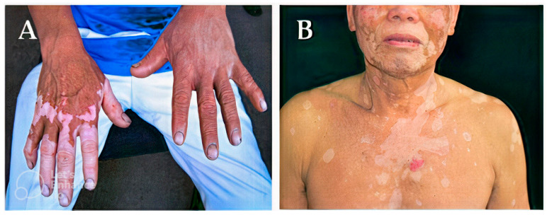
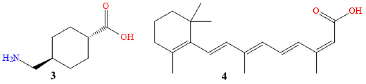
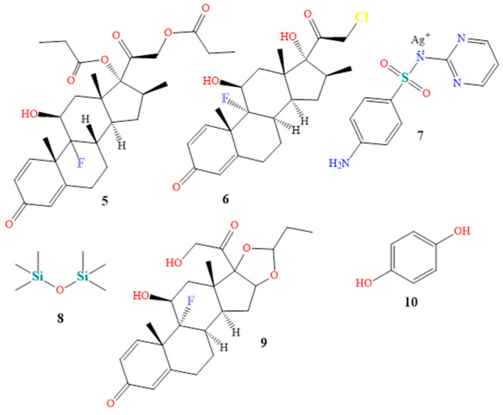
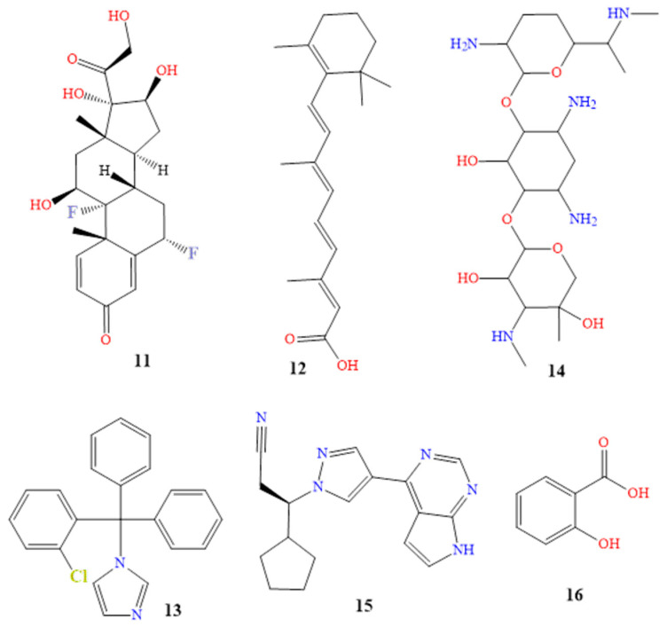
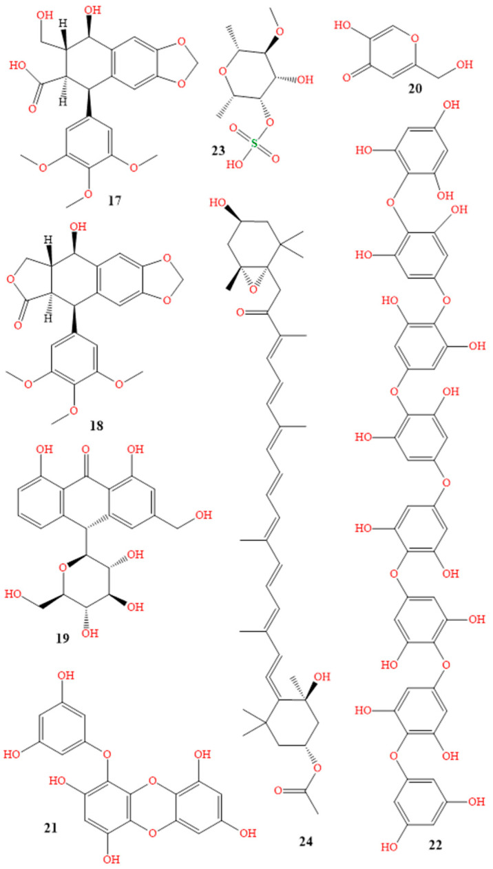
Human skin pigmentation and melanin synthesis are incredibly variable, and are impacted by genetics, UV exposure, and some drugs. Patients' physical appearance, psychological health, and social functioning are all impacted by a sizable number of skin conditions that cause pigmentary abnormalities. Hyperpigmentation, where pigment appears to overflow, and hypopigmentation, where pigment is reduced, are the two major classifications of skin pigmentation. Albinism, melasma, vitiligo, Addison's disease, and post-inflammatory hyperpigmentation, which can be brought on by eczema, acne vulgaris, and drug interactions, are the most common skin pigmentation disorders in clinical practice. Anti-inflammatory medications, antioxidants, and medications that inhibit tyrosinase, which prevents the production of melanin, are all possible treatments for pigmentation problems. Skin pigmentation can be treated orally and topically with medications, herbal remedies, and cosmetic products, but a doctor should always be consulted before beginning any new medicine or treatment plan. This review article explores the numerous types of pigmentation problems, their causes, and treatments, as well as the 25 plants, 4 marine species, and 17 topical and oral medications now on the market that have been clinically tested to treat skin diseases.

Keywords: depigmentation; hyperpigmentation; hypopigmentation; melanin; skin lightening; skin pigmentation; tyrosinase inhibitors; vitiligo.


Conflict of interest statement

The authors declare no conflict of interest.

Figures

Figure 3
Figure 3
Hypopigmentation cases (A [22], B [23]) and hyperpigmentation cases (C,D [24]). (A) Reproduced with permission from Joo-Heung Lee, Cho-Rok Kim, Dong-Youn Lee, Photodermatology, Photoimmunology & Photomedicine; published by John Wiley and Sons, 2011. (B) Reproduced with permission from Jeremy P Hill, Jonathan M Batchelor, The BMJ; published by BMJ Publishing Group Ltd., 2017. (C,D) Reproduced with permission from Narumol Silpa-archa, Indermeet Kohli, Suteeraporn Chaowattanapanit, Henry W. Lim, Iltefat Hamzavi, Journal of the American Academy of Dermatology; published by Elsevier, 2017.
Figure 1
Figure 1
Schematic representation of the melanin synthesis pathway.
Figure 2
Figure 2
Pigmentation from prolonged UV exposure formed: (A) stratum corneum, (B) epidermis, (C) dermis, (D) melanocyte cell, (E) tyrosinase and tyrosine, (F) melanin, and (G) keratinocyte cell.
Figure 4
Figure 4
The mechanisms involved in some types of hypopigmentation.
Figure 5
Figure 5
Depigmentation cases [27,28]. (A) Reproduced with permission from Louise McMichael, the BMJ; published by BMJ Publishing Group Ltd., 2012. (B) Reproduced with permission from Jing Jing, Xiao-Yong Man, the BMJ; published by BMJ Publishing Group Ltd., 2021.
Figure 6
Figure 6
Chemical structures of tranexamic acid (3), and isotretinoin (4).
Figure 7
Figure 7
Chemical structures of betamethasone (5), clobetasol (6), silver sulfadiazine (7), dimethicone (8), triamcinolone (9), and hydroquinone (10).
Figure 8
Figure 8
Chemical structures of fluocinolone (11), tretinoin (12), clotrimazole (13), gentamicin (14), ruxolitinib (15) and salicylic acid (16).
Figure 9
Figure 9
Structures of podophyllum resin (17), podofilox (18), aloin (19), kojic acid (20) phlorotannins (21), octaphlorethol A (22), fucoidan (23), and fucoxanthin (24).

References

    1. Del Bino S., Duval C., Bernerd F. Clinical and biological characterization of skin pigmentation diversity and its consequences on UV impact. Int. J. Mol. Sci. 2018;19:2668. doi: 10.3390/ijms19092668. - DOI - PMC - PubMed
    1. Martin A.R., Lin M., Granka J.M., Myrick J.W., Liu X., Sockell A., Atkinson E.G., Werely C.J., Möller M., Sandhu M.S., et al. An unexpectedly complex architecture for skin pigmentation in Africans. Cell. 2017;171:1340–1353. doi: 10.1016/j.cell.2017.11.015. - DOI - PMC - PubMed
    1. Nasti T.H., Timares L. MC 1R, Eumelanin and Pheomelanin: Their role in determining the susceptibility to skin cancer. Photochem. Photobiol. 2015;91:188–200. doi: 10.1111/php.12335. - DOI - PMC - PubMed
    1. Solano F. Photoprotection and skin pigmentation: Melanin-related molecules and some other new agents obtained from natural sources. Molecules. 2020;25:1537. doi: 10.3390/molecules25071537. - DOI - PMC - PubMed
    1. Polidori C., Jorge A., Ornosa C. Eumelanin and pheomelanin are predominant pigments in bumblebee (Apidae: Bombus) pubescence. PeerJ. 2017;5:e3300–e3321. doi: 10.7717/peerj.3300. - DOI - PMC - PubMed

LinkOut - more resources