. 2023 Jun 7;12(6):814.
doi: 10.3390/pathogens12060814.

CLEAR Strategy Inhibited HSV Proliferation Using Viral Vectors Delivered CRISPR-Cas9

Affiliations

CLEAR Strategy Inhibited HSV Proliferation Using Viral Vectors Delivered CRISPR-Cas9

Min Ying et al. Pathogens. .

Abstract

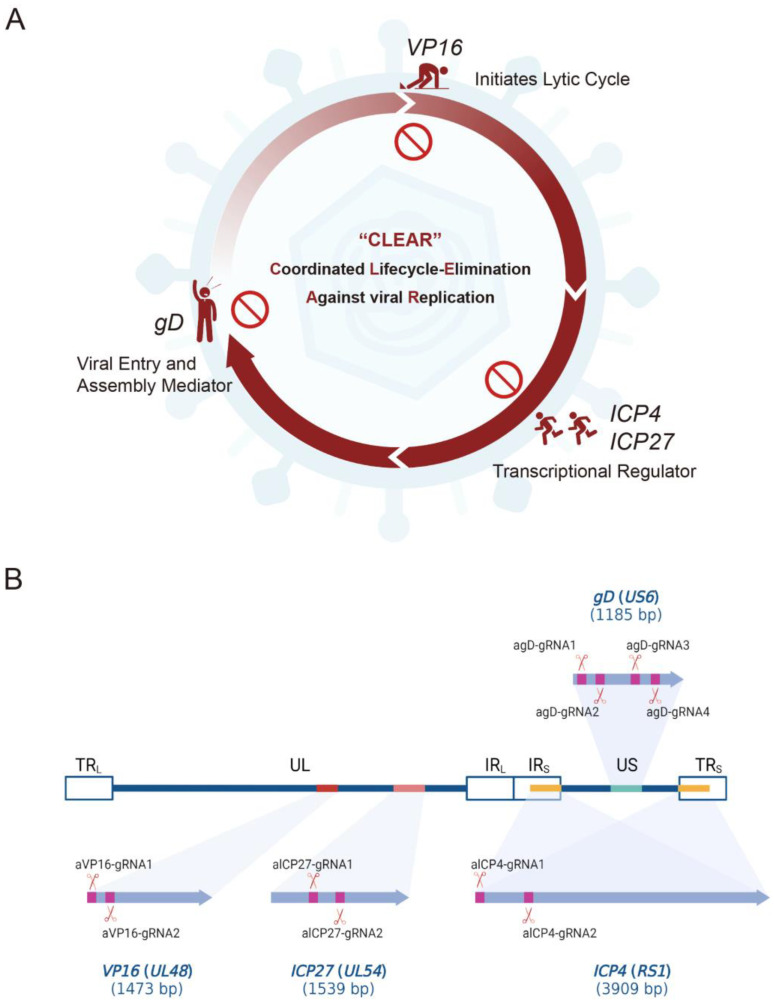
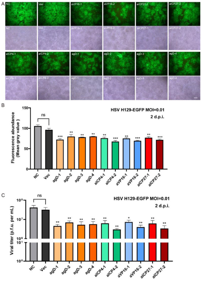
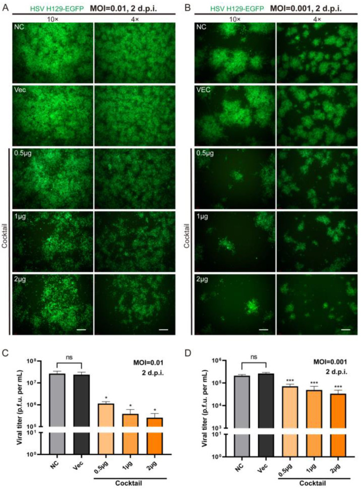
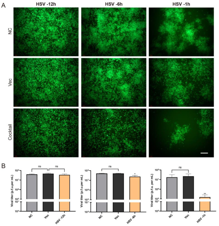
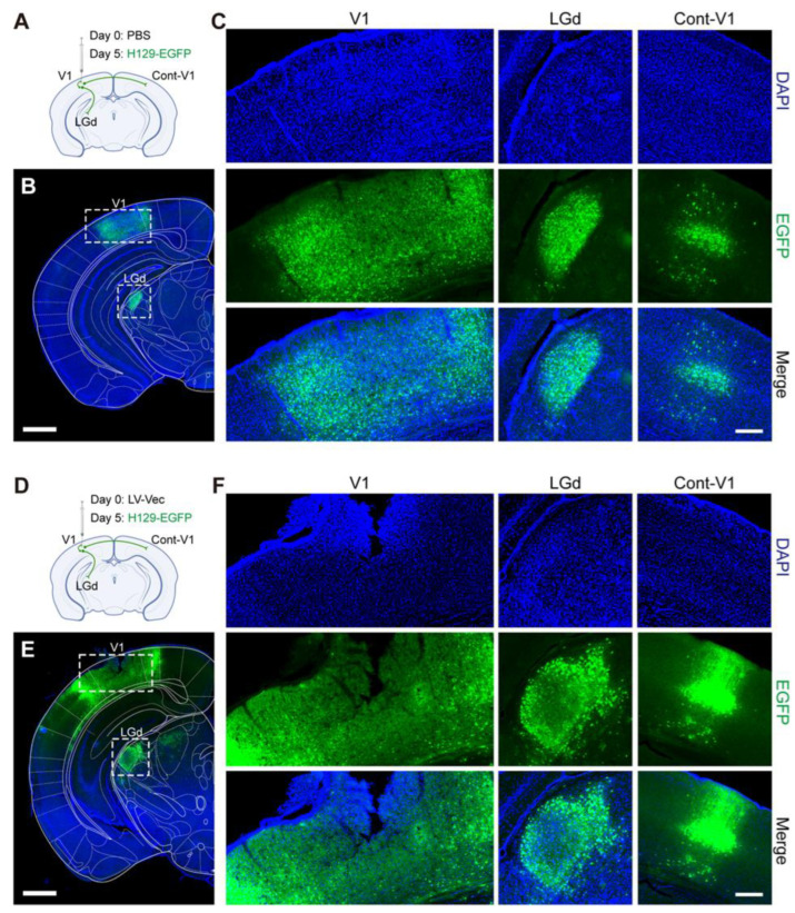
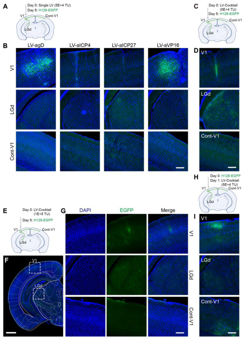
Herpes simplex virus type 1 (HSV-1) is a leading cause of encephalitis and infectious blindness. The commonly used clinical therapeutic drugs are nucleoside analogues such as acyclovir. However, current drugs for HSV cannot eliminate the latent virus or viral reactivation. Therefore, the development of new treatment strategies against latent HSV has become an urgent need. To comprehensively suppress the proliferation of HSV, we designed the CLEAR strategy (coordinated lifecycle elimination against viral replication). VP16, ICP27, ICP4, and gD-which are crucial genes that perform significant functions in different stages of the HSV infection lifecycle-were selected as targeting sites based on CRISPR-Cas9 editing system. In vitro and in vivo investigations revealed that genome editing by VP16, ICP27, ICP4 or gD single gene targeting could effectively inhibit HSV replication. Moreover, the combined administration method (termed "Cocktail") showed superior effects compared to single gene editing, which resulted in the greatest decrease in viral proliferation. Lentivirus-delivered CRISPR-Cas9/gRNA editing could effectively block HSV replication. The CLEAR strategy may provide new insights into the potential treatment of refractory HSV-1-associated diseases, particularly when conventional approaches have encountered resistance.

Keywords: CRISPR-Cas9; ICP27; ICP4; VP16; gD; gRNA; herpes simplex virus; lentivirus.


Conflict of interest statement

The authors declare no conflict of interest.

Figures

Figure 1
Figure 1
The schematic diagram of the CLEAR (coordinated lifecycle elimination against viral replication) strategy and the designed gRNAs targeting essential HSV1 genes. (A) Schematic representation of the CLEAR strategy. VP16, ICP27, ICP4, and gD play different essential roles in the different stages of the HSV proliferation cycle. (B) The schematics of 10 designed gRNAs and their targeting and cleavage sites. The process involves utilizing lentiviral vectors to deliver SpCas9 and viral gene-targeting gRNAs, ultimately aiming to eliminate key viral genes and inhibit HSV replication.
Figure 2
Figure 2
Gene verification and editing activity analysis of constructed LV-Cas9-gRNA plasmids. (A) PCR confirmation of the correct gRNA cassette integration into the lentiCRISPR-v2 vector. Lane 1, molecular size marker (M); Lane 2, negative control; Lane 3, 1885 nucleotide fragments consistent with the parental vector amplified product (mesoplasma florum L1 gene) size; Lanes 4–13, the right amplified products of constructed lentiviral plasmids consistent with correct the insertion of the gRNAs cassette. (B,C) Mutation detection in targeted cells by T7E1 assay. BHK-21 cells were transfected with 2 μg of LV-Vec or LV-Cas9-gRNA plasmids and subsequently infected with H129-EGFP at MOI = 0.01 24 h later. Cells were harvested 48 h post infection, and lysates were used to amplify target loci. Amplified products were denatured/annealed and digested with T7 Endonuclease I according to the recommended protocol. The resulting fragments were consistent with the expectation. The corresponding fragment sizes, as marked by the red arrows, were as follows: Con, 514 bp = 335 bp + 179 bp; agD-1, 803 bp = 436 bp + 367 bp; agD-2, 803 bp = 410 bp + 393 bp; agD-3, 837 bp = 418 bp + 419 bp; agD-4, 837 bp = 467 bp + 370 bp; aICP4-1, 910 bp = 545 bp + 365 bp; aICP4-2, 910 bp = 521 bp + 389 bp; aVP16-1, 900 bp = 517 bp + 383 bp; aVP16-2, 900 bp = 497 bp + 403 bp; aICP27-1, 897 bp = 521 bp + 376 bp; aICP27-2, 897 bp = 481 bp + 416 bp.
Figure 3
Figure 3
The inhibitory effect of CRISPR single gene editing on HSV replication in vitro. (A) Cas9-gRNA editing inhibited HSV replication in BHK-21 cells. Different Cas9/gRNA plasmids were transfected into BHK-21 cells, and cells were infected with HSV H129-EGFP 24 h later. Cell fluorescence images were collected 48 h after HSV infection (MOI = 0.01). The bright field images show the total number of cells, complete confluence, and inter-group consistency of the results. Scale bar = 200 μm; (B) the fluorescence abundance (average gray value) of EGFP expression at 48 h post-infection. (C) The viral titers of HSV were determined. Statistical values were presented as mean ± SEM. Significant differences were expressed by the p value. ns, no significant difference; * p < 0.05, ** p < 0.01, *** p < 0.001 (sgRNA versus Vec).
Figure 4
Figure 4
The efficacy of the CLEAR strategy in inhibiting HSV replication in vitro. (A) Fluorescence images of cells 48 h post cocktail transfection following HSV infection (MOI = 0.01), with both 4× and 10× magnification images. (B) Fluorescence images of cells 48 h post-cocktail transfection following HSV infection (MOI = 0.001), with both 4× and 10× magnification images. Images at 4×; scale bar = 500 μm. Images at 10×; scale bar = 200 μm. (C) The viral titers of HSV infection (MOI = 0.01) 48 h after cocktail transfection; cocktail versus Vec. ns, no significant difference; * p < 0.05. (D) The viral titers of HSV infection (MOI = 0.001) 48 h after cocktail transfection; cocktail versus Vec. ns, no significant difference; *** p < 0.001. The statistically significant reduction in viral titers after cocktail transfection further supported the efficacy of the CLEAR strategy in inhibiting HSV replication in vitro.
Figure 5
Figure 5
The protection effect of the CLEAR strategy after HSV1 pre-infection. (A) The cell fluorescence images of cocktail transfection groups at 1 h, 6 h, and 12 h after HSV infection. Scale bar = 200 μm. (B) The viral titers of gRNA cocktail editing groups at 1 h, 6 h, and 12 h after HSV infection (MOI = 0.01). ns, no significant difference; * p < 0.05; ** p < 0.01.
Figure 6
Figure 6
The control mice received either PBS or LV-Vec showed strong HSV1 replication and spread after viral challenge. (A) Diagram illustrating circuit connections and the infusion of PBS and H129-EGFP. The dots on the green lines represent the cell body of the neuron, and the end of the green lines represent the axon terminals. V1, primary visual area; LGd, dorsal part of the lateral geniculate complex; Cont-V1, contra-lateral V1. PBS (500 nL) and H129-EGFP (5 × 10² PFU) were injected 5 days apart (n = 3). (B) The dissemination of HSV1 H129-EGFP viral infection in the PBS control group. Scale bar = 1 mm. (C) The fluorescence distribution of EGFP at the injection site of V1 and downstream areas of LGd and contra-lateral V1. Scale bar = 200 μm. (D) Diagram illustrating circuit connections and the viral infusion of LV-Vec and H129-EGFP. LV vector control (LV-Vec, 5 × 10⁴ TU) and H129-EGFP (5 × 10² PFU) were injected 5 days apart. (E) The dissemination of HSV1 H129-EGFP viral infection in the LV-Vec control group. Scale bar = 1 mm. (F) The fluorescence distribution of EGFP at the injection site of V1 and downstream areas of LGd and contra-lateral V1. Scale bar = 200 μm.
Figure 7
Figure 7
Lentiviral vectors delivering gene-editing elements effectively cleared HSV1 H129 in vivo. We designed four experimental groups: (A,B) four individual LV treatments, (C,D) a mixed treatment group (LV-Cocktail) with equal amounts of each single LV, (EG) a double-concentration mixed treatment group (LV-Cocktail), and (H,I) a group inoculated with HSV first and then injected with LV. (A) Diagram illustrating circuit connections and the viral infusion of HSV1 H129-EGFP and the single LV vector. Single LV (5 × 10⁴ TU) and H129-EGFP (5 × 10² PFU) were injected 5 days apart (n = 3). (B) The fluorescence distribution of EGFP at the injection site of V1 and downstream areas of LGd and contra-lateral V1 via the single administrated LV. Scale bar = 200 μm. (C) Diagram illustrating circuit connections and the viral infusion of HSV1 H129-EGFP and low doses of the LV-Cocktail. LV-Cocktail (5 × 10⁴ TU) and H129-EGFP (5 × 10² PFU) were injected 5 days apart. (D) The fluorescence distribution of EGFP at the injection site of V1 and downstream areas of LGd and contra-lateral V1. Scale bar = 200 μm. (E) Diagram illustrating circuit connections and the viral infusion of HSV1 H129-EGFP and high-dose LV-Cocktail. LV-Cocktail (1 × 10⁵ TU) and H129-EGFP (5 × 10² PFU) were injected 5 days apart. (F) The dissemination of HSV1 H129-EGFP after clearance by high-dose LV vectors 5 days later; scale bar = 1 mm. (G) The fluorescence distribution of EGFP at the injection site of V1 and downstream areas of LGd and contra-lateral V1 via high dose of administrated LV. Scale bar = 200 μm. (H) Diagram illustrating circuit connections and the viral infusion of HSV1 H129-EGFP and the LV-Cocktail vector (LV-Cocktail administration after first infection with HSV). H129-EGFP (5 × 10² PFU) and LV (5 × 10⁴ TU) were injected 1 day apart. (I) The fluorescence distribution of EGFP at the injection site of V1 and downstream areas of LGd and contra-lateral V1. Scale bar = 200 μm.

Similar articles

Cited by

References

    1. James C., Harfouche M., Welton N.J., Turner K.M., Abu-Raddad L.J., Gottlieb S.L., Looker K.J. Herpes simplex virus: Global infection prevalence and incidence estimates, 2016. Bull. World Health Organ. 2020;98:315–329. doi: 10.2471/BLT.19.237149. - DOI - PMC - PubMed
    1. Kukhanova M.K., Korovina A.N., Kochetkov S.N. Human herpes simplex virus: Life cycle and development of inhibitors. Biochemistry. 2014;79:1635–1652. doi: 10.1134/S0006297914130124. - DOI - PubMed
    1. Klysik K., Pietraszek A., Karewicz A., Nowakowska M. Acyclovir in the Treatment of Herpes Viruses—A Review. Curr. Med. Chem. 2020;27:4118–4137. doi: 10.2174/0929867325666180309105519. - DOI - PubMed
    1. Khadr L., Harfouche M., Omori R., Schwarzer G., Chemaitelly H., Abu-Raddad L.J. The Epidemiology of Herpes Simplex Virus Type 1 in Asia: Systematic Review, Meta-analyses, and Meta-regressions. Clin. Infect. Dis. 2019;68:757–772. doi: 10.1093/cid/ciy562. - DOI - PMC - PubMed
    1. Baringer J.R., Swoveland P. Recovery of herpes-simplex virus from human trigeminal ganglions. N. Engl. J. Med. 1973;288:648–650. doi: 10.1056/NEJM197303292881303. - DOI - PubMed

LinkOut - more resources